| Feel the fetlock joint and sesamoids for heat or swelling. |
The pictures and diagrams in the anatomy portion of Section A focus on the
major bones and joints of the front and hind limbs. Refer to these pictures for
basic anatomy and definitions. When describing a bone or joint, realize that
there can be up to three different names for each structure.
To simplify, the descriptions used from this point on will use the more commonly used terms.
Diagnosing Lameness: There are many different reasons for a horse to become lame. Because of this, even the professionals struggle at times with a diagnosis. However, there are a few basic tips, that if understood, can help even the most casual horse owner become fairly adept at detecting a lameness problem.
Tip #1 - Studies show that most lameness problems are associated with the foot. Therefore, the foot should be the first area of focus. Realize that 60-65% of the horse’s body weight is carried on the front legs, resulting in more problems occurring in the front legs and feet.
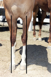
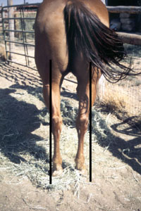
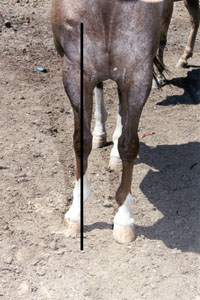
Tip #2 - Conformation (how the horse is put together) plays a major role in lameness problems. Initially, the following observations should be made:
|
Figure 1
|
|
Figure 2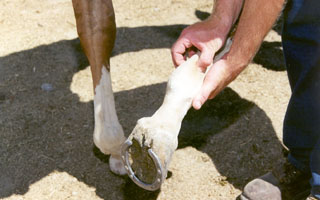
|
|
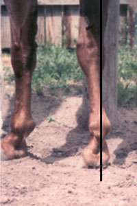
Tip #3 - Observe the horse at rest. Look and feel for heat/swelling or the way the horse is holding a suspect limb. (See figures 1-2.) These little hints will help narrow the possibilities.
Tip #4 - Observe the horse at exercise. Initially, this can be done first at walking speeds, then at a trot or gallop, if necessary. Choose a hard surface to maneuver the horse in straight lines and then in circles.
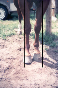
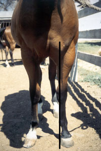
Tip #5 - Observe the horse from behind, side, and front. As each foot hits the ground, listen for differences in sound on impact. Look for interfering, overreaching, and scalping. (See figure 3.)
Figure 3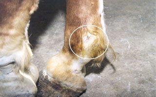
|
|
Tip #6 - A horse that is lame on a
front limb will:
- Realize that the horse uses its head to help take weight off the lame limb.
Tip #7 - A horse that is lame on a
hind limb will:
Figure 4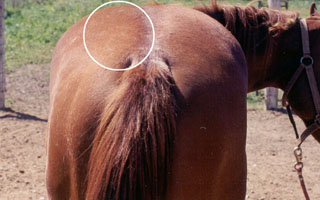
|
|
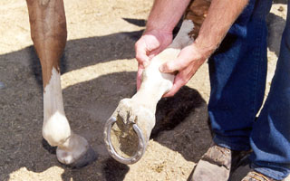
Tip #8 - After a specific limb is determined to be the problem, various other techniques can be utilized to help localize the specific area that is causing the lameness. These include the following:
 |
| Fig. 5 |
* Realize that experience and training are essential to identify animals that are only slightly lame.
Figure 6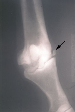
|
|
Figure 7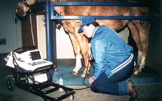
|
|
Treatment:
Depending on the specific cause of the lameness, one or all of the following may be necessary:|
|
||||||
|
|
|
||||||
|