F218
Eye Diseases and Problems

More Cat Info

BUY THIS MANUAL NOW and have access to this article and 100's of others just like it!

View some of the 15+ Video clips found in the Feline Manual


basic anatomy and terms | nasolacrimal duct system | proptosis of the eyeball | glaucoma | entropion | ectropion | bilateral protrusion of the third eyelid | prolapsed gland of the third eyelid | blepharitis | tumors of the eyelid | conjunctivitis | abrasions, lacerations | keratitis | lenticular sclerosis | cataract | luxation and subluxation of the lens | anterior uveitis | feline generalized retinal atrophy | hypertensive retinopathy | feline central retinal degeneration | cancers | eye medications


Introduction: It is important to examine the eyes of a pet on a routine basis. Repeated examination allows one to become familiar with the normal appearance of the eye, so any abnormalities will be noticed immediately. Signs of an eye problem vary tremendously and may include cloudiness, tearing, squinting, discharge, redness, blinking, swelling, an increase in blood vessels, or changes in the size or shape of the pupil. Cats may paw at the eye or rub it in an attempt to relieve irritation and itching. Any change in the eye or surrounding tissue may signal a problem and should be a cause for concern.

Many different problems can result in the same set of disease signs, so diagnosis cannot be made by clinical signs alone. Physical examination and special tests are needed to properly identify the cause of the problem. Tests may include a fluorescein dye test, a test for tear production called a Schirmer test, tests to measure pressure within the eyeball, and ocular examination with different types of lenses. The eye may be dilated to allow for proper visualization of the back of the eye.

Eye problems should be brought to the attention of a veterinarian immediately. Prompt diagnosis and treatment can prevent further eye problems that can lead to loss of sight. In addition, changes in the eyes may be a sign of whole body disease. By immediately identifying and reporting any changes, diseases can be diagnosed early and treatment can begin.

Basic Anatomy and Terms: The eye is protected by upper and lower lids, as well as a third eyelid, called the nictitating membrane. Glands which produce tears are located under the lids. The front portion of the eye itself is covered with a thin, clear covering called the cornea. The remainder of the eye is covered with dense, white tissue, the sclera. The margin of the cornea and the sclera is called the limbus. The episclera is the outside surface of the sclera. The conjunctiva is the tissue which reflects from the inside of the eyelids onto the globe. Glands which produce tears are also located in the conjunctiva.

The iris is the colored portion of the eye; the black open space in the iris is the pupil. Behind the pupil is the lens. The lens is attached to the ciliary body. The back of the eye is covered with a layer of tissue called the retina. The inside of the globe is divided by the lens into two chambers. The forward, smaller chamber is called the anterior chamber. This chamber is filled with a clear fluid called aqueous humor. This fluid is produced by the ciliary body and nourishes the eye while helping to maintain its shape. This fluid is continually produced and drained from the eye. Drainage occurs at the iridocorneal angle, also called the drainage or filtration angle. The larger chamber behind the lens is called the posterior chamber. This chamber is also filled with a clear, thick, gelatinous substance called aqueous humor.

Glossary of Eye Terms:

  1. Anterior chamber - The space in the eye bounded in front by the cornea and behind by the iris. It is filled with aqueous humor.
  2. Anterior uvea - The front portion of the uvea, made up of the iris and ciliary body.
  3. Anterior uveitis - Inflammation of the iris and ciliary body.
  4. Anti-inflammatory - A medication that reduces inflammation.
  5. Aqueous humor - The clear, watery fluid which fills the eye.
  6. Blepharitis - Inflammation of the eyelids.
  7. Blepharospasm - Spasm of the muscles that control the eyelids.
  8. Cataract - An opacity of the lens or its capsule, or both.
  9. Choroid - The part of the vascular layer of the eye (uvea) located at the back of the eye. It helps to nourish the retina. It is the posterior uvea.
  10. Ciliary body - The part of the vascular layer of the eye (uvea) located between the iris and the choroid.
  11. Conjunctiva - The mucous membrane lining the back of the eyelids and the front of the eye, except for the cornea.
  12. Conjunctivitis - Inflammation of the conjunctiva.
  13. Cornea - The transparent portion of the outer layer of the eye, which allows light to enter the eye. It attaches to the sclera.
  14. Corneal pigmentation - The deposition of pigment in the cornea, usually as a response to chronic irritation.
  15. Corneal ulceration - An open lesion or sore on the surface of the cornea.
  16. Dry eye - See keratoconjunctivitis sicca.
  17. Enucleation - Removal of the eye.
  18. Epiphora - The abnormal flow of tears over the face.
  19. Fibrous - Composed of cells that form tough connective tissue.
  20. Fluorescein dye - A special dye that is applied to the cornea that will outline ulcerations in the cornea. The dye will not penetrate intact cornea or conjunctiva, but will adhere to and outline areas of ulceration.
  21. Glaucoma - Increased ocular pressure resulting from altered flow of aqueous humor.
  22. Globe - The eyeball.
  23. Intraocular - Within the eyeball.
  24. Iris - The colored, circular portion of the eye located behind the cornea. It is part of the uvea.
  25. Keratoplasty - Corneal grafting used as a repair technique.
  26. Keratectomy - Removal of part of the superficial layers of the cornea.
  27. Keratitis - Inflammation of the cornea.
  28. Keratoconjunctivitis sicca (KCS) - A condition of the eye caused by lack of proper tear production that results in inflammation of the cornea and conjunctiva. Also called dry eye. It is rare in the cat.
  29. Lacrimal apparatus - The structures in the eye responsible for tear production.
  30. Lenticular sclerosis - A gradual hardening and thickening of the lens that accompanies aging. This does not interfere with vision.
  31. Lens - A transparent structure suspended in the front portion of the eye. It refracts light that passes through the cornea on its way to the retina.
  32. Lens luxation - Displacement of the lens.
  33. Miosis - Contraction of the pupil so that the pupil becomes small.
  34. Miotic - A medication that causes the pupil to become small.
  35. Mucoid - This is a general term for types of secretions that are made up of mucous materials.
  36. Mucopurulent - A secretion or exudate that is made up of pus and mucous.
  37. Mydriatic - A medication that causes the pupil to become large.
  38. Mydriasis - A dilated pupil.
  39. Nasolacrimal - The system of the eye responsible for proper drainage of tears out of the eye and into the inside of the nose.
  40. Nictitans (nictitating membrane) - The third eyelid.
  41. Ophthalmoscope - A device for studying the interior of the eyeball.
  42. Optic nerve - The nerve that originates from the retina and carries light signals to the brain.
  43. Pigmentation - Any abnormal coloration of an area.
  44. Posterior uvea - The portion of the uvea made up of the choroid.
  45. Posterior uveitis - Inflammation of the choroid.
  46. Pupil - The circular opening in the center of the iris through which light passes into the eye.
  47. Primary - The first, or foremost. Used in this case to describe problems that occur first and may be followed by other, secondary problems.
  48. Proptosis - Prolapse of the eye.
  49. Retina - The innermost coat of the eye, formed of cells sensitive to light.
  50. Retinal degeneration - A progressive, pathological change that interferes with the function of the retina.
  51. Schirmer tear test - A method of measuring tear production using small strips of absorbent paper placed into the lower corner of the eye.
  52. Sclera - This is a layer of dense connective tissue that supports the eye. It is the white portion of the eye that connects to the cornea. The cornea and sclera make up the fibrous tunic of the eye.
  53. Secondary - A problem or disease which occurs after the first or primary disease or problem.
  54. Tonometry - The measurement of intraocular pressure.
  55. Topical - Medication that is applied locally on an external portion of the eye.
  56. Uvea - The portion of the eye which contains many blood vessels. It is comprised of the iris, ciliary body, and choroid. It is called the vascular tunic.
  57. Uveitis - Inflammation of the uvea.
  58. Vascular - Containing blood vessels.
  59. Vascularization - The formation of new blood vessels in a part of the body.


  1. Anterior chamber     
  2. Cornea
  3. Suspensory lig.
  4. Ciliary body
  5. Sclera
  6. Choroid
  7. Vitreous chamber
  8. Optic disc
  9. Retina
  10. Lacrimal gland
  11. Eyelid
  12. Pupil
  13. Iris
  14. Lens


Copyrighted graphic used by permission from Anatomy of Domestic Animals, Sudz Publishing (email: sudzpub@mac.com)


BUY THIS MANUAL NOW and have access to this article and 100's of others just like it!

Nasolacrimal Duct System and Lacrimal System

Introduction: The lacrimal and nasolacrimal duct system function together to ensure that the eye is continuously bathed in protective tears and that the tears are removed from the eye. The lacrimal system is responsible for the production of tears. Glands in the eye produce tears that act to lubricate the eye and cornea, protect the eye from environmental debris, supply nutrients to the eye, and wash away irritants. Both the lacrimal glands and the gland of the third eyelid contribute to tear production. After the tears protect and bath the eye, they are drained via the nasolacrimal system. This duct system is responsible for drainage of tears from the inner corner of the eye into the inside of the nose. Health of the cornea and conjunctiva depends on the continuous production of tears and their uninterrupted removal from the eye.

If there is a problem with any part of the system, drainage of tears from the eye may be impeded. If the tears do not drain into the duct system, they will spill over onto the face, creating a condition called epiphora. Signs include wet facial skin and a reddish, rusty stain to the skin and hair near the inner corners of the eyes.

Problems Associated with the Nasolacrimal System

  1. Absence of the Puncta: There can by many different causes for obstructions in the nasolacrimal duct system. Blockage will occur if the openings to the ducts, called the puncta, are absent or closed. Cats may be born with closed or absent puncta. Diagnosis is based on physical examination and specialized tests. Tests may include the use of fluorescein dye to observe the movement of tears, a nasolacrimal flush, and passage of a nasolacrimal catheter. If a potentially functional duct system exists, treatment involves opening the puncta with surgery. Once the duct is created or opened, a catheter can be placed for one to two weeks to ensure it remains open. The eye is also treated simultaneously with topical medication to reduce inflammation and prevent infection.
     
  2. Obstruction of the Puncta or Ducts: Other causes of obstructions may include inflammation that closes down any part of the duct system. This obstruction can be caused by conjunctivitis or by physical irritants such as sand, plant stems, and other foreign objects. The puncta may also be blocked because of herpesvirus infections. Diagnosis is based on ocular examination and appropriate testing. Treatment of an obstructed nasolacrimal duct would include antibiotic, antiviral, and/or anti-inflammatory medical therapy, removal of any foreign objects, and flushing of the duct system. Additional causes of obstruction may include space-occupying tumors. Treatment would depend upon the type and location of the tumor and may include surgical removal.
     
  3. Epiphora: Some cats, such as certain Persian blood lines, have facial anatomy that predisposes them to have tears drain down the front of the face. These cats may have one or more of several conditions contributing to improper drainage of tears. These conditions can include the hair near the eyes acting as a wick to pull the tears onto the face, absence of the nasolacrimal puncta, obstruction of any part of the nasolacrimal duct system, excessive tear production caused by irritation or infection, inverted lower eyelids (entropion), or a prominent eye with a very small area for tears to pool. Typically, tears pool together in the inner corner of the eye before entering the duct system. Any cat with a flattened face and large, prominent eyes has a diminished area for the tears to pool, resulting in spillage over the lower lid.

    Diagnosis is based on physical examination and testing. Treatment may include surgery to correct underlying anatomical abnormalities, topical medications such as antibiotics, oral medications, and daily cleaning of the face and hair. Many cats respond favorably to treatment with oral antibiotics, but the epiphora returns when the medication is stopped. Medical control requires long term continuous or intermittent treatment.

    Prognosis depends on the cause of the epiphora and elected treatment protocol. If the condition is not associated with any other ocular problems, it may be considered a cosmetic issue and can be controlled with daily cleaning of the affected area.

Problems with the Eye and Associated Structures:

Globe or Eyeball

  1. Proptosis of the Eyeball: Proptosis literally means "falling forward." In this case, the eyeball pops out if the socket. This condition typically occurs following facial trauma, such as an automobile accident or a cat fight. Breeds of cats with flat faces, such as certain Persian blood lines, are prone to this occurrence. Because of their facial anatomy, minor trauma can result in the eyeball popping out of the socket. The problem can, however, occur in any animal if the trauma is great enough. In rare cases, a tumor behind the eyeball causes it to be pushed out of the socket.

    This condition is easily identified because the globe is out of the socket and pushed in front of the eyelids. This results in swelling, cloudiness, eyelid entrapment, and inflammation of all associated structures. Other accompanying signs may include an ulcerated or dried out (desiccated) cornea, abnormal pupil size, hemorrhage of the eye or in the conjunctiva, and rupture of the globe. If the optic nerve is cut or compressed, blindness may result. Depending on the cause, other signs of facial trauma, such as fractures, bruising, and bleeding, may be evident. The animal may also be in shock or have other signs of trauma to the face or body.

    To prevent blindness, emergency treatment should be started as rapidly as possible. The cornea should be lubricated immediately to prevent further damage. The animal must be assessed, and shock and other traumatic damage treated as necessary. If the patient is stable, the globe should be returned to its socket surgically. This is most easily done under sedation or anesthesia. This is followed by medical treatment to reduce swelling and prevent or treat infection. The globe cannot be repositioned if it is severely damaged, infected, or desiccated, or if the optic nerve is severed; these eyes are usually removed. Follow-up treatment involves both whole body (systemic) and topical medication with antibiotics and corticosteroids as indicated by the condition of the eye and the patient.

    Prognosis depends on the time from onset until treatment, along with the amount of damage to the eye and surrounding structures. Potential outcomes include a permanently dilated pupil, decreased tear production, desiccation of the cornea, and turning of the eye to a position in the upper, outer corner of the eye (dorsolateral strabismus). Blindness in both eyes can possibly result if enough damage is done to the optic chiasm, where both optic nerves connect to each other before traveling to the brain.

    Some cats will improve over time, others will not. Most eyes can be saved; however, some may need to be removed. Many cats will not regain sight. Permanent blindness can result if the optic nerve is injured, treatment is delayed, or there is too much damage or swelling to replace the globe. Removal of the globe may be necessary if pain or damage to the eye is present.
     
  2. Glaucoma: Glaucoma is defined as an abnormal increase in pressure inside the eyeball (intraocular pressure or IOP). It is caused by impairment to the drainage of aqueous humor. Aqueous humor, the fluid that nourishes the eye, is continually produced and drained from the eye. It is drained around the perimeter of the eye at an area called the iridocorneal angle. This area is bounded in the front at the location where the cornea meets the sclera and behind at the location where the lens meets the musculature of the ciliary body. If this area is blocked, drainage is impeded. If drainage is impeded, fluid and pressure build in the eye, damaging internal structures. Damage to the retina and optic nerve can result in permanent loss of vision.

    Glaucoma can either exist as the primary problem, or it may be secondary to other diseases, such as luxation of the lens, uveitis, neoplasia, or bleeding in the eye. The condition may be acute or chronic. Primary glaucoma can be an inherited disease in cats and is thought to occur in the Siamese breed more than other feline breeds.
     
    Figure #2: The eye on the left shows the typical signs of glaucoma: cloudiness, corneal edema, and a dilated pupil.

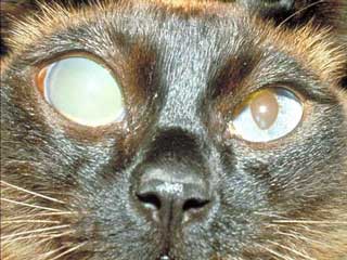
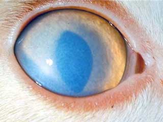
    Acute glaucoma has a rapid onset. The rapid increase in intraocular pressure results in an extremely painful eye. The pain can cause squinting (blepharospasm) and epiphora. The cat may resist examination, refuse to eat, be depressed, and even cry out from the pain. In addition, ocular discharge, redness, and cloudiness may be noticed.

    Diagnosis is based upon examination of the eye and measurement of the pressure in the globe. Upon examination, the glaucomatous eye may have cloudiness, corneal edema, redness, and engorgement of the blood vessels in the episclera. The pupil may be dilated and unresponsive to light. The globe may be enlarged and firm to the touch. Accompanying problems such as uveitis, a luxated lens, or neoplasia may be noted. Specific eye examination may show a closure of the normal drainage area of the eye. The blood vessels in the retina may be compressed by the increase in pressure. Measurement of the intraocular pressure will show it to be elevated. Definitive diagnosis is based on measurement of an elevated intraocular pressure. Acute glaucoma can very rapidly lead to blindness. Total, irreversible blindness can occur in as little as 24-48 hours.


    Treatment of acute glaucoma is considered an emergency. Rapid treatment may prevent permanent blindness. If the glaucoma is secondary to an underlying cause, treatment of the primary cause may result in resolution of the glaucoma. Medical treatment is initially used for treatment of primary glaucoma. Medical treatment may include the use of diuretics, such as mannitol, and topical medications to reduce aqueous production. Drugs can be applied directly to the eye to reduce aqueous humor production, including carbonic anhydrase inhibitors such as dichlorphenamide, and B-adrenergic antagonists such as timolol maleate. Other drugs that help to open drainage and enhance the outflow of aqueous humor may be tried. These include pilocarpine and demecarium bromide. Cats do not tolerate topical eye medications for treatment of glaucoma as well as dogs do, and the dose of topical drug used must be lowered for cats. Some irritation to the eye may still result from the medications even at the lowered dose.

    If medications are insufficient to lower the intraocular pressure, surgical treatment is needed. Surgery may be done to reduce the production of aqueous humor and to create drainage. Laser treatment may be used. A combination of medical and surgical therapies may be needed for long-term control of the problem. It is important to note that if only one eye is involved, both eyes should be medicated. If treatment is not successful and blindness results, removal of the eye may be recommended to eliminate the pain associated with the condition. If complete eye removal (enucleation) is unacceptable, the contents of the eye may be removed (eviscerated) and an insert placed inside the eye (intraocular prosthesis) to maintain its form.

    Chronic glaucoma occurs if the signs of acute glaucoma go unnoticed or if therapy is not effective. The signs may include all of those noticed with acute glaucoma, although to a lesser degree. The most obvious sign is the enlarged globe. In addition, corneal ulceration, lens luxation, cataracts, and keratitis may occur. Ocular examination will show degeneration of the retina and the head of the optic nerve. The animal is typically blind in the affected eye. Diagnosis is based on the physical and ocular examination.

    Treatment is aimed at reducing the pain of chronic glaucoma. The blindness is not reversible. The medical and surgical treatments described for the control of acute glaucoma may be used. Removal of the eye may be needed to control pain. If only one eye is affected, the normal eye should be examined and monitored for any signs of glaucoma. Preventative (prophylactic) treatment of the unaffected eye should be started if indicated by ocular examination.

    Prognosis for treatment of glaucoma depends on the underlying cause and the time between onset of disease and treatment. Treatment to maintain or restore vision will be unsuccessful in acute glaucoma cases unless initiated within hours of onset. Chronic glaucoma patients may be blind before treatment begins. Secondary glaucoma cases can resolve following successful treatment of the primary cause. This can occur with glaucoma secondary to lens luxation.

BUY THIS MANUAL NOW and have access to this article and 100's of others just like it!

Eyelids

Introduction: Cats have three eyelids. The upper and lower lids help to protect the eye from the environment, distribute tears over the entire eye surface, and control the amount of light that enters the eye. The third eyelid is located in the inner corner of the eye and sweeps across the eye as it closes. It functions to protect and lubricate the eye. It has its own set of tear glands that produce lubricating tears for the entire eye.

Problems with the lids can result in pain, swelling, redness, excessive tearing, and abnormal drainage from the eye. Cats with lid pain may rub their eyes, paw at their faces, squint, and show other signs of pain. Problems with the lids can lead to additional problems with closely associated structures such as the cornea, conjunctiva, and nasolacrimal drainage system of the eye. The majority of lid problems that are common in dogs are uncommon in cats.

  1. Entropion: This is a condition characterized by an inward turning of either upper or lower eyelids. Occasionally both upper and lower eyelids may be affected. The inward turning is called inversion. The turned-in lids can result in irritation and abrasion of the cornea caused by contact with either the eyelid itself or hairs on the eyelid. This can lead to corneal ulceration, which will interfere with vision. Severe, untreated entropion can lead to perforation of the cornea and permanent blindness.

    Diagnosis is based on physical examination. Treatment involves eye medications such as antibiotics and anti-inflammatory medications to treat underlying causes of squinting and spasm. Temporary sutures may be placed to evert the lid and decrease irritation to the eye. Once underlying causes are treated, those cases that continue to have entropion are treated with surgical techniques to roll the eyelid out and reduce irritation.
     
  2. Ectropion: This is a rolling out of the eyelids. The outward turning is called eversion. This results in exposure of the conjunctiva. Exposure of the conjunctiva can result in chronic inflammation and disease of the conjunctiva and cornea. Chronic changes in the cornea caused by ectropion can lead to loss of vision.

    Signs of ectropion include the visible outward rolling of the eyelid with exposure of the conjunctiva. Associated conditions include drainage from the eye, recurrent infections of the eye, and irritation and inflammation of the conjunctiva. Drainage may be mucoid (looks like mucus) or mucopurulent (looks like mucus and pus) and can be accompanied by staining of the face. Cats with a history of ectropion may continually get conjunctivitis, eye irritation, or foreign bodies in their eyes.

    Treatment includes applying lubricants and protectants to the eye and conjunctiva and treating infections with appropriate antibiotics. Daily cleaning of the eyes and face are part of this therapy. If conservative therapy does not control the irritation and infection, definitive therapy includes corrective surgery to reduce or lift the eyelid.

    Prognosis is good with appropriate therapy. Cats treated with non-surgical techniques must be monitored for signs of worsening disease or secondary infections.
     
  3. Bilateral Protrusion of the Third Eyelid: Also called "haws," this condition is most common in cats under 3 years of age. An underlying digestive tract infection is usually present, such as tapeworms or viral infections.

    Treatment with topical epinephrine or phenylephrine administered 3-4 times per day may help relieve clinical signs. Most cases will spontaneously recover within several weeks. Additional treatment options include topical medications to reduce swelling and irritation, along with a surgical procedure where the gland is sutured back in place.

    If replacement is not successful, the gland can be removed, but removal is neither the best nor the first method of treatment. Removal of the gland can result in lack of tear formation that leads to development of corneal and conjunctival problems. Surgical removal is therefore avoided if it is at all possible to replace the gland. Topical therapy with anti-inflammatory eye medication, such as corticosteroids, may be helpful to reduce swelling before surgery and is important as an aid to help the eye heal after surgery. Prognosis is good with prompt surgical repair.
     
  4. Prolapsed Gland of the Third Eyelid: Also called "cherry eye," this condition is characterized by the prolapse of the gland that typically sits beneath the third eyelid. If the attachment of the gland weakens, the gland can move upward and out of its normal position. It will then become visible as a red, oval bulge protruding just behind the edge of the third eyelid. It is often called a cherry eye due to the visible appearance of the red bulge. It may affect one or both eyes.
     
    Figure #3: This picture shows a protrusion of the third eyelid identified by the white arrow.

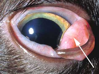
    The condition is diagnosed on physical examination by the appearance of the red, round to oval mass coming up over the edge of the third eyelid. Accompanying signs can include squinting, repeated blinking, increased tearing, and a reddening of the conjunctiva.

    Treatment includes topical medications to reduce swelling and irritation, along with a surgical procedure where the gland is sutured back in place. If replacement is not successful, the gland can be removed, but removal is neither the best nor the first method of treatment. Removal of the gland can result in lack of tear formation that leads to development of corneal and conjunctival problems. Surgical removal is therefore avoided if it is at all possible to replace the gland. Topical therapy with anti-inflammatory eye medication, such as corticosteroids, may be helpful to reduce swelling before surgery and is important as an aid to help the eye heal after surgery.
     

  5. Blepharitis: Blepharitis is inflammation of the eyelids. It can be caused by any condition that results in inflammation, including allergies, parasites, immune-mediated disease, endocrine disease, trauma, neoplasia, nutritional deficiencies, and infection. Infection can be caused by bacteria, viruses, or fungi.

    All cats can suffer from blepharitis. It may involve one or more lids, and be localized to one spot or involve large areas of the involved lids. Signs may include eyelid spasm, squinting, pain, ocular discharge, discoloration of the lids, hair loss surrounding the lids, redness, crusting, abscesses, pustules, and swelling. There may be concurrent irritation and scarring of the cornea, and redness and swelling of the conjunctiva.

    Diagnosis is based on physical examination and specialized tests. A Schirmer tear test, cultures and sensitivities, cytology, skin scrapings, and biopsies may be performed. Underlying conditions such as fungal infections, endocrine disease, parasitic skin infections, bacterial skin infections, and allergies must be diagnosed and eliminated. Once a diagnosis is reached, treatment is designed to eliminate the underlying cause and remove the inflammation from the eyelid.

    Treatment may involve medications such as antibiotics, antifungals, and anti-inflammatories, along with topical medications that are instilled into the eye and on the lids. Topical therapies may include appropriate antibiotics, corticosteroids, and combination antibiotic/anti-inflammatory medications.

    Prognosis depends on the cause of the inflammation. Some cases respond rapidly to treatment. Other cases that are caused by whole body (systemic) disease may respond only after treatment of the animal’s underlying problem and may require management for the lifetime of the animal. Proper diagnosis is critical for long-term resolution of blepharitis.
     
  6. Tumors of the Eyelid: Growths can occur on the eyelids. The growths most commonly occur in older animals. Most masses and growths that occur on the eyelids are benign. Those that are malignant tend to invade immediate tissues, but do not typically spread (metastasize) to other places. Squamous cell carcinoma is the most frequently seen tumor in cats.

    Cats with eyelid masses will show various signs depending on the location of the mass. Blepharitis is common. If the mass contacts the conjunctiva, irritation and conjunctivitis will result, leading to signs that include epiphora, ocular discharge, and squinting. If the mass contacts the cornea, corneal irritation and infection may result. This may cause squinting, epiphora, photosensitivity, and other signs of keratitis.

    Diagnosis is based upon physical examination. Treatment involves removal of the mass by appropriate surgical techniques, which will vary depending on the size and location of the mass. Reconstructive eyelid surgery may be needed. Prognosis is typically good, but depends on the size and type of the mass. The majority of masses are benign and removing the mass will usually cure the problem. If removal is not complete, many masses will return. Most malignant masses are also controlled by surgical removal.

BUY THIS MANUAL NOW and have access to this article and 100's of others just like it!

Conjunctiva

Introduction: The conjunctiva is the membrane that lines the inside of the eyelids and the third eyelid, and covers the outside of the sclera. The conjunctiva is a mucous membrane with an excellent blood supply. It connects the lids to the globe and contains specialized glands. These glands produce the inner layer of the tear film. Problems affecting the conjunctiva may be limited to only the conjunctiva or may involve other portions of the eye. Conjunctival inflammation or disease may also signal illness that affects the entire body. It is important to recognize whether disease processes are limited to the conjunctiva, extend to other parts of the eye, or signal whole body (systemic) disease.

  1. Conjunctivitis: Inflammation of the conjunctiva is termed conjunctivitis. Any cat can suffer from conjunctivitis, but those cats with either underlying allergies or other eye diseases tend to get conjunctivitis more frequently than cats with normal eyes and no immunological problems. Conjunctivitis may be primary or secondary to other problems.

    Primary conjunctivitis can be caused by immune-mediated diseases, allergies, foreign objects in the conjunctiva, or environmental irritants (dust, chemicals). The majority of the cases of conjunctivitis are secondary to other eye problems such as lid malformations or nasolacrimal diseases. Others occur following glaucoma, cancer, uveitis, corneal inflammation, or systemic immune-mediated diseases. Cats are also prone to having upper respiratory infections of Chlamydia, herpesvirus, and Mycoplasma that can lead to conjunctivitis.

    Signs of conjunctivitis may include spasm of the eyelids, swelling and redness of the conjunctiva, and discharge from the eyes. The discharge may be clear, mucoid, or mucopurulent. Cats with conjunctivitis may be sensitive to and avoid the light. They may respond to the conjunctival irritation by pawing at the eyes and rubbing the face and eyes.
     
    Figure #4: This picture shows a cat with severe conjunctivitis.

    Conjunctivitis is diagnosed on physical examination. Additional tests are performed to identify other eye problems and to rule out other eye diseases that can cause conjunctivitis. These include a Schirmer tear test, fluorescein dye, intraocular pressure test, bacterial culture and sensitivity, and conjunctival cytology. Steps are taken to identify any underlying or accompanying disease situations that contribute to the conjunctival inflammation.

    Treatment involves both medicating the conjunctiva and treating any underlying or secondary problems. Depending on the cause of the conjunctivitis, treatment may include topical medications for inflammation and infection, eye washes to remove discharge, lubricants to add moisture to the eye, and medications to control infection and inflammation. Topical therapy may include antibiotic agents, corticosteroids, or combination medications. All discharge should be flushed from the eye before treatment is attempted to allow the medications to contact the surface of the eye and the conjunctiva.

    Prognosis depends on the underlying cause and severity of the condition. Simple bacterial conjunctivitis is typically very responsive to treatment with the appropriate antibacterial medications. Secondary conjunctivitis may not respond until the underlying cause is identified and treated. Some secondary conjunctivitis problems may be controlled, but not totally eliminated.

Cornea

Introduction: The cornea is the outer, transparent layer of the front of the eye. It protects the eye while still allowing light to pass through. The cornea is protected by a layer of tears and by continuously replacing its superficial cells. It lacks blood vessels, and so does not heal easily. Any disease process or insult to the cornea can result in cloudiness (edema), swelling, or pigmentation, which in turn may lead to loss of vision. Corneal irritation or inflammation is extremely painful. It is critical to treat any corneal problem quickly.

Figure #5: This picture shows a cat with corneal edema. Notice how the surface of the eye appears cloudy.
  1. Abrasions, Lacerations: Any animal can suffer from a traumatic corneal abrasion, scratch, or laceration. Mild injuries can result in a superficial abrasion. Deeper wounds are identified as penetrating or perforating. A penetrating laceration involves the cornea, but does not pass through it. A perforation goes entirely through the cornea.

    If wounds are caused by a penetrating or perforating object, the object may still be in the cornea. Wounds can be caused by any object that can contact the eye. Common injuries occur as a result of cats being hit by pellets from guns, scratched by other cats, and running through twigs and branches. Blunt trauma from being hit by a car or running into an immovable object can also lead to lacerations of the cornea. Cats that engage in fights with other cats, and cats that climb through bushes or trees are at an increased risk for corneal abrasions and cuts.

    Signs of a corneal wound include pain and inflammation of the eye. The eyelids may be kept closed and the cat may resist examination. The wound may be visible on the cornea, along with swelling, cloudiness, and hemorrhage. Facial trauma may also be evident. Diagnosis is made by history and physical examination. An immediate diagnosis can be made if the object that caused the wound is still in the eye. Additional testing is done to determine the severity and extent of the wound, assess vision, and identify trauma to other portions of the eye and face. Bacterial culture and sensitivity may be necessary. A specific test called a fluorescein dye test is performed using special stain to ascertain if the cornea is intact or if it has been perforated.

    Treatment depends on the severity, depth, and age of the wound. Care should be taken to apply only minimal pressure to the face and eye to prevent rupture of the eyeball. Any foreign body should be flushed out, removed with a dampened cotton-tipped applicator, removed with forceps, or surgically removed from the eye. Superficial wounds can be treated with topical medications to prevent infections and calm the eye. Topical eye medications include antibiotics and atropine. In addition, an Elizabethan collar or similar collar is used to prevent the cat from scratching the eye as it heals. Activity should be restricted and the cat kept indoors to avoid further injury.

    Some lacerations may require additional therapy with a soft contact lens. This type of therapy is usually available only through a specialist. Extensive corneal lacerations and corneal penetrations require surgical repair. Surgical repair will also be needed for those injuries that contain a foreign body that is not easily removed. All corneal wounds should be rechecked at specific intervals to monitor healing. Superficial wounds can be checked at 3 day intervals; deep wounds should be checked daily for the first few days.

    Prognosis depends on the extent of the wound and the time elapsed between injury and veterinary treatment. The deeper the wound, the poorer the prognosis. Most superficial corneal abrasions and wounds heal quickly and satisfactorily. Deeper wounds or those created by blunt trauma may result in permanent loss of vision, inflammation, and pain. The full extent of the damage caused by the wound may not be apparent for days to weeks following the injury, so re-examination is necessary.
     
  2. Keratitis: Keratitis refers to inflammation of the cornea. Signs may include swelling (edema), cloudiness, and pigmentation. Ulcerative keratitis is inflammation accompanied by ulceration of the surface of the cornea (a corneal ulcer). This is a very common condition in cats, and all breeds may be affected. Breeds with flat faces may have greater risk of ulcerative keratitis. Ulcerative keratitis can be caused by any condition that disrupts the layers of the cornea, including trauma, entropion, ectropion, trichiasis, keratoconjunctivitis sicca, damage to the nerves of the face, burns caused by chemicals and cleaners, and diseases of the immune system. Infection by bacterial or fungal organisms is uncommon, but may occur secondary to other irritations and injuries.

    Ulcerative keratitis
    is a very painful condition. Signs of disease include tearing (lacrimation), spasms of the lids, pawing at the face, squinting, spilling of tears onto the face (epiphora), avoidance of sunlight (photophobia), redness, and discharge from the eye. One or more defects in the cornea may be visible and are referred to as corneal ulcer(s). Some ulcerative keratitis cases have a mushy, gelatinous appearance as the cornea breaks down due to excessive enzyme production. Chronic ulcers may show vascularization, scarring, pigmentation, and swelling. Healing ulcers may have a hazy, white appearance. Fluorescein dye testing (shown below) helps to determine the depth, severity, and number of ulcers.
     
    Figure #6: Small circular areas with finger-like projections are areas of corneal ulceration highlighted by fluorescein dye.

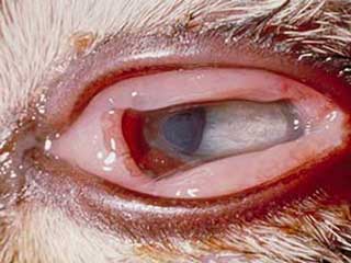
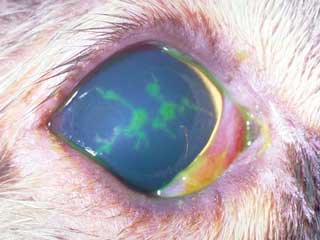
    Diagnosis is based on ocular examination and fluorescein dye testing. If needed, specific examination of the interior of the eye and cytology can also aid in the diagnosis. Other tests, such as a Schirmer tear test, are performed to rule out additional or contributory eye diseases.

    Treatment involves elimination of the cause, along with specific treatment for the ulceration and inflammation. Underlying eye problems, such as keratoconjunctivitis sicca, entropion, or ectropion, should be treated appropriately. Treatment of the ulcer may include topical antibiotics to prevent infection, topical atropine to control pain, specific medications to control fungal or viral infections, and if indicated, specific medications to prevent collagen breakdown.

    Some ulcers are treated with protective contact lenses. Others may require surgery to trim (debride) the ulcer edges. Additional surgical procedures include punctuate keratotomy, conjunctival flaps, and flaps created from the third eyelid. Proper use of an Elizabethan collar or similar device will help prevent the animal from scratching the healing ulcer. Eyes should be rechecked at approximately 3-day intervals; those with deep ulcers should be rechecked daily until satisfactory healing is observed.

BUY THIS MANUAL NOW and have access to this article and 100's of others just like it!

Lens

Introduction: The lens focuses light waves that come through the pupil. It is held in place by small suspensory ligaments called lens zonules that attach the lens to the ciliary body. The ciliary body can contract and relax, thereby changing the shape of the lens. The changing shape of the lens allows it to properly focus light waves from different distances onto the retina.

  1. Lenticular Sclerosis: Lenticular sclerosis, also called nuclear sclerosis, is a normal aging change in older cats. It is commonly seen in cats 10-12 years of age or older. As the cat ages, the lens thickens and becomes less flexible. This thickening creates haziness in the eye that can be confused with cataract formation. Light will pass through a sclerotic lens; the cloudiness does not obscure vision. Nuclear sclerosis can be differentiated from a true cataract on ocular examination. No treatment is necessary.
     
  2. Cataract: Cataracts refer to either opacities within a lens or an entire lens that is opaque. The term cataract means "to break down," referring to the loss of normal architecture of the fibers in the lens or its capsule, resulting in an increase of fluid in the lens and loss of transparency. Cataracts can be hereditary. In cats with hereditary cataracts, the cataracts may appear at any age and are often seen in younger kittens. The type of cataract and age at which it appears is breed specific and can be associated with other eye abnormalities.

    Although most cataracts in cats are hereditary, other factors can contribute to the formation of cataracts. These can include metabolic diseases, trauma to the globe, exposure to toxins, radiation therapy performed near the eyes, certain amino acid deficiencies, and inflammatory diseases such as uveitis.

    Although cataracts can be seen early in their development, most owners do not notice any signs of cataracts until the animal loses sight. Owners may not notice the loss of vision if the cat gradually adapts to the loss, but will be aware of a problem when total blindness results. Not all cataracts develop to the point where they cause blindness. Diagnosis is made by examination of the eyes. Cataracts can be classified by location in the lens and degree of development. Examination of the retina should accompany any examination for cataracts.

    Treatment is usually reserved for cataracts that cause blindness. There is no effective medical therapy. A limited number of cataracts will spontaneously resolve. Spontaneous cataract resorption is an unpredictable process that may or may not result in improved vision. It is not advisable to delay treatment while attempting to wait for spontaneous resorption. Treatment involves surgical removal of the lens. The lens can be replaced with a prosthetic lens if desired. Although several techniques can be used, phacoemulsification is commonly used today. This technique involves the use of a small ultrasonic probe that is placed into the eye. It shatters the cataract and then removes the broken-down debris by suction.

    Prognosis depends on the severity of the cataract, the cause of the cataract, and the time between identification and surgical removal. Success rates with phacoemulsification are greater than 90%. The best prognosis is associated with hereditary cataracts treated early in their course. The worst prognosis is associated with long standing cataracts that are found in conjunction with other ocular problems, such as uveitis or degeneration of the retina.
     
  3. Luxation and Subluxation of the Lens: This occurs if the lens separates from the zonules that hold it in position. A partial separation results in a subluxation of the lens, leaving the lens either in or near its normal position. A total luxation results from a complete rupture of the zonules and total separation of the lens. The lens will then displace either into the anterior or posterior chamber. This condition can occur either by itself or secondary to intraocular problems including uveitis, glaucoma, neoplasia, or trauma.

    Signs of lens subluxation or luxation vary depending on the degree of lens movement and its location. A subluxation may not result in signs that are noticed by the owner. Luxation may result in a painful eye with photophobia, edema of the cornea, cloudiness of the cornea, and redness. Examination of the eye reveals changes in the depth of the anterior chamber of the eye, iridodonesis (the iris moves rapidly or trembles when the eye is moved), and an aphakic crescent. An aphakic crescent is a crescent-shaped area of the pupil that is lacking the lens. This occurs because the luxated lens is no longer sitting directly behind the pupil, but has shifted its position. Other eye conditions, such as glaucoma or uveitis, may be present.

    Diagnosis is based on ocular examination. The anterior chamber will be abnormally deep or shallow and the aphakic crescent (an area where the lens is not present) may be visible. Additional tests for ocular disease may be performed, including pressure tests for glaucoma and dilation of the eye to check for other abnormalities. Because lens luxation can occur in both eyes, the "normal" eye should also be examined.

    Treatment depends on the location and displacement of the lens. Subluxations may not require any treatment, except for continual monitoring. Subluxated lenses can progress to total luxations or contribute to glaucoma. Lenses that have displaced into the posterior chamber of the eye may be managed with topical medications, such as 0.125% demecarium bromide, that keep the pupil contracted.

    The majority of luxated lens are treated with surgical removal. If surgery is delayed, topical anti-inflammatory medications, such as 0.1% dexamethasone, are indicated, along with medications to reduce intraocular pressure and prevent glaucoma. These include dichlorphenamide, a carbonic anhydrase inhibitor. Surgery should be performed as soon as possible to prevent loss of vision. If accompanying glaucoma has resulted in blindness or a neoplasia is present, treatment may involve removal of the eye.

    Prognosis depends on the degree of displacement and location of the lens, accompanying ocular disease, and time between onset of the problem and treatment. The best prognosis is reserved for those cases that are treated early after onset with surgery and do not have accompanying glaucoma. The poorest prognosis accompanies those cases with glaucoma and a delay between onset and treatment.

BUY THIS MANUAL NOW and have access to this article and 100's of others just like it!

Uvea

Introduction: The uvea is a very vascular structure that is critical for the maintenance of a healthy eye. It is a pigmented, vascular tunic that sits between the outer fibrous layer of the eye (cornea and sclera) and the inner nervous layer (retina).  It is comprised of 3 connected portions - the iris, the ciliary body, and the choroid. The anterior uvea is made up of the iris and the ciliary body. The posterior uvea, located towards the back of the eye, is comprised of the choroid. The iris controls the amount of light that enters the eye. The ciliary body controls the focus of the lens, produces aqueous humor, and helps regulate intraocular pressure. The anterior uvea acts as a blood-aqueous barrier and prevents unwanted particles from the bloodstream from entering the aqueous humor. The choroid provides nourishment to the retina. Most diseases of the choroid are linked to disease of the retina. Because the uvea is highly vascular, it is very reactive to changes in the body and is easily inflamed.

Inflammation of the uvea is called uveitis. Specifically, inflammation of the iris and ciliary body is termed anterior uveitis. Posterior uveitis refers to inflammation of the choroid. Inflammation may be limited to only the anterior or posterior uvea, or involve both portions. Inflammation of the uvea allows particles to cross the blood-aqueous barrier and enter the aqueous humor. This causes an inflammatory response in the aqueous which can lead to a loss of vision.

Anterior uveitis: Inflammation of the anterior uvea may occur with several disease conditions seen in the eye. The cause may be external to the eye and include trauma. In addition, anterior uveitis may follow corneal injuries and corneal ulceration. Anterior uveitis can also be caused by infectious agents such as viruses, bacteria, fungi, and parasites. These infections can be limited to the eye or cause disease throughout the body. Common infections in the cat include feline leukemia virus (FeLV), feline infectious peritonitis (FIP), and toxoplasmosis. In addition, certain immune-mediated diseases, cancers of the eye, and exposure to some toxins may lead to anterior uveitis.

Signs of anterior uveitis may include pain, redness of the conjunctiva, corneal edema, red blood cells or white cells in the anterior chamber, epiphora, spasm of the eyelids, and aversion to light. Other ocular changes are specific to anterior uveitis. These include constriction of the pupil, a change in the color of the iris (often darker), swelling of the iris, and enlargement and engorgement of deep blood vessels located in the ciliary body. In addition, inflammatory cells and pigment may clump together and adhere to the cornea, forming small, visible masses called "keratic precipitates." As particles form or flood into the inflamed area, the aqueous humor becomes turbid. This condition is referred to as "aqueous flare." In chronic cases, the iris may actually adhere to the lens, "fixing" the pupil in an unmovable position. This results in a distorted and immobile pupil. Finally, anterior uveitis can be associated with other serious eye problems, including lens luxation, cataracts, ulcerative keratitis, and glaucoma.

Figure #7: This picture shows an eye inflicted with uveitis.

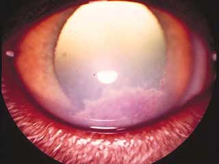
Diagnosis is based upon complete physical and ocular examination. A thorough eye examination includes measurement of the intraocular pressure. The pressure is typically decreased with uveitis. Additional testing may include blood tests, urinalysis, and radiographs to search for the underlying cause of the disease.

Treatment is aimed at reducing the inflammation of the uvea while determining and eliminating the underlying cause. Anti-inflammatory agents including corticosteroids (1% prednisolone acetate, 0.1% dexamethasone) and non-steroidal anti-inflammatories (flurbiprofen) can be applied topically. Topical medications that dilate the pupil (atropine) are also used. In some cases, corticosteroids can be injected under the conjunctiva or administered systemically. Those patients that do not respond to corticosteroids can be treated with other immunosuppressive drugs, such as azathioprine. Treatment may be altered depending on the cause of the uveitis and the systemic illnesses that are present. Treated animals should be re-examined within one week following the initial treatment and re-evaluated every few weeks.

The animal’s prognosis depends on the severity of the uveitis at the time of treatment and the underlying cause of the uveitis. Early, aggressive treatment of the uveitis and the initiating cause is necessary to prevent secondary problems. Prognosis is good if the underlying cause is identified and eliminated, and appropriate eye therapy is instituted. If uveitis is left untreated, glaucoma, lens luxation, and blindness can result. Successful treatment can involve several months of continual medication and follow-up examinations.

* All of the eye pictures were used with permission from Colorado State University Ophthalmology Service.

Retina

Introduction: The retina is the back portion of the eye. It is considered the "film" that records the visual images that come through the cornea and are focused by the lens. The cells of the retina receive the light images. These cells are of two main types, rods and cones. The rods are sensitive to dim light and the cones are sensitive to bright light. The rods are useful at dusk, while the cones perceive images in the day and help distinguish colors.

The rods and cones translate the light images into chemical messages which affect adjacent nerve fibers. The retinal nerve fibers converge together at an area called the optic disc to form the optic nerve. The messages travel as nerve impulses along the optic nerve to the brain where they are again converted into visual images. Any type of damage to the retina will result in interruption of this process and cause loss of vision.

Feline Generalized Retinal Atrophy: If the retina degenerates or atrophies, messages from the eye will no longer reach the brain and blindness will result. Conditions such as vitamin A or taurine deficiency can cause this problem. Retinal atrophy has also been associated with certain breeds of cats (Persians, and Siamese), suggesting that it can be an inherited problem.

With retinal atrophy, early cases may not show many signs. As the retina’s cells degenerate, the cat loses vision. Cats will often lose night vision first, so the animals may have difficulty in dimly lit rooms or at night. This problem can go unnoticed by an owner that leaves on indoor lights until retiring for the evening. Eventually, the retinal atrophy will progress and total blindness will result. An owner may be aware of dilated pupils or a shine from the back of the dilated eye, or may notice nothing until the animal has totally lost vision.

Diagnosis is based upon ocular examination. Pupils will be dilated and not responsive to light. The retinal area will be hyperreflective and retinal examination will show degeneration of the retina and optic nerve, along with changes in the retinal blood vessels and pigmentation. A test called an electroretinography can be performed to measure electrical activity in the retina. The most common treatment options include dietary changes that increase the levels of vitamin A and taurine in the diet.

Hypertensive Retinopathy: Disease and dysfunction of the retina in older cats due to high blood pressure (hypertension) is another cause of blindness. High blood pressure leads to rupture of delicate blood vessels in the retina with separation (detachment) of the retina occurring in some cases. Blood pressure can be difficult to accurately measure in cats. Hyperthyroidism (see page F815) is a common cause of high blood pressure in older cats. If the blood pressure can be effectively lowered with specific medication or proper treatment of hyperthyroidism, some vision may be regained.

Feline Central Retinal Degeneration: This problem is associated with inadequate levels of taurine in the diet. Unlike retinal atrophy, cats that have central retinal degeneration do not have any vision problems and their pupils do not remain dilated. There is some thought that this problem will progress into generalized retinal atrophy. Treatment involves feeding taurine.

Cancers

Introduction: Cancers may develop in any structure in the eye. They can be found in any age or breed of cat, but are generally more common in older animals. A cancer may be primary and arise from tissue in the eye, or be secondary to a cancer somewhere else in the body. Cancers located anywhere else in the body may migrate (metastasize) to the eye. Tumors may be localized nodules or locally invasive. In addition to occupying space, tumors may cause infection and inflammation of involved tissues. Common eye tumors include adenomas, adenocarcinomas, papillomas, melanomas, histiocytomas, squamous cell carcinomas, and fibromas. Tumors are more likely to be found on the eyelids than any other part of the eye.

A mass in or near the eye will cause signs that reflect the involved area of the eye. For example, a mass on the eyelid can cause signs of blepharitis and conjunctivitis. A mass located on the retina may cause blindness and a dilated pupil. Signs associated with eye disease are not specific to one diagnosis, so ocular examination and diagnostic tests are necessary for proper diagnosis. Diagnostic tests may include special examination, biopsy, histopathology, and MRI testing. Microscopic examination (histopathology) will allow differentiation between inflammation, benign tumors, and malignant tumors. Treatment depends on the type and location of the growth and may include surgical removal, chemotherapy, and removal of the eye. Prognosis depends on the location and type of growth. Early identification and removal of malignant tumors will increase the chances of a successful outcome and reduce tumor spread.

BUY THIS MANUAL NOW and have access to this article and 100's of others just like it!

Eye Medications: Eye medications can be delivered by several methods. Topical medications are applied directly to the eye surface. The topical medications may be available as eye drops and ointments. This method of administration is appropriate for both hospital and home treatment of eye diseases in cats. In addition, veterinarians may administer medications via injection into the eye. Common sites for these injections are subconjunctival (beneath the conjunctiva), retrobulbar (behind the eye), or intraocular (into the eyeball). In addition, diseases of the eye may be treated with medications that are given directly to the cat, either by mouth or by injection. Finally, eye diseases may not be limited to the eyes; they may be a sign of disease that is affecting the entire body. In this case, the veterinarian will prescribe medication to treat the primary illness, as well as to control the problems in the eyes. The following tables list commonly used eye medications.

CLASSIFICATION/USE/INDICATION MEDICATION SPECIFIC USE NOTES CONTRA-INDICATION (IF ANY)
EYE RINSES

USE: Clean, rinse, flush

INDICATIONS: Clear mucus before instilling medications, remove debris from eye

Sterile, buffered isotonic solutions containing sodium chloride, sodium citrate, sodium phosphate    
Combinations of water, boric acid, zinc sulfate    
       
EYE LUBRICANTS

 USE: Lubricate, prevent eye irritation, relieve dryness

INDICATIONS: Keratoconjunctivitis sicca, whenever general anesthesia is used, keratitis, ectropion

Pilocarpine Irritating, can cause conjunctivitis and worsen uveitis, not commonly used Can affect respiration and cardiac function
Polyvinylpyrrolidone    
Polyvinyl alcohol    
Methylcellulose    
Ethylene glycol polymers    
Refined petrolatum    
Refined lanolin    
Refined peanut oil    
       
MUCOLYTICS

USE: Prevent collagen break-down, break up mucus

INDICATIONS: "Melting" corneal ulcers, chronic conjunctivitis, keratoconjunctivitis

Acetylcysteine   Very expensive
Autologous plasma   Sometimes used in place of acetylcysteine
       
ANESTHETICS

USE: Topical pain relief

INDICATIONS: Minor surgery, eye examination, diagnostic procedures, preoperative evaluation of entropion, removal of foreign bodies

Proparacaine 0.5%   Never use therapeutically. May cause corneal irritation.
Tetracaine HCl 0.5%*  
       
ANTIBIOTICS (SINGLE)

USES: Preparation for an intraocular procedure. 

Treatment of infection (if possible, select specific agent for microbe; if testing is not possible, broad spectrum or combination antibiotic is preferred.) 

Preventive pre and/or post-procedure.

INDICATIONS: Treat susceptible infections contributing to uveitis, conjunctivitis, blepharitis, keratitis, keratoconjunctivitis sicca. 

Control secondary bacterial infections in conditions such as proptosis of the globe, entropion, ectropion, corneal ulcer, corneal abrasion.

Chloramphenicol 0.5% solution and 1% ointment Susceptible bacteria may include Staphlococcus, Streptococcus spp, Corynebacterium, Hemophillis spp, Moraxella, Chlamydia, Mycoplasma spp.  
Gentamicin 0.3% solution and 0.3% ointment Susceptible bacteria may include Staphylococcus, Corynebacterium, Pseudomonas, Proteus spp, Escherichia coli, Hemophillis, Enterobacter, Moraxella  
Tetracycline 1% solution and 1% ointment Susceptible bacteria may include Staphylococcus, Corynebacterium spp,, Hemophillis spp, Moraxella, Chlamydia, Mycoplasma spp  
Tobramycin 0.3% solution and 0.3% ointment Susceptible bacteria may include Pseudomonas, Proteus spp, Escherichia coli, Hemophillis, Enterobacter, Moraxella, Staphylococcus  
Bacitracin 500 U/g ointment Susceptible bacteria may include Staphlococcus, Streptococcus, Corynebacterium spp  
Chlortetracycline 1% ointment    
Erythromycin 0.5% ointment    
Neomycin 0.35% ointment Susceptible bacteria may include Staphylococcus, Corynebacterium spp, Hemophillis spp, Moraxella, Enterobacter, Mycoplasma  
       
ANTIBIOTICS (COMBINATION)

USE: Same as single antibiotic 

When more than one type of microbe is present or when testing for specific identification is not possible.

INDICATIONS: Same as single antibiotic

Neomycin sulfate, Polymyxin B sulfate solution and ointments    
Neomycin sulfate, Polymyxin B sulfate, gramacidin solution Preferable drug for broad spectrum coverage without culture/sensitivity  
Neomycin sulfate, Polymyxin B sulfate, Bacitracin ointment Preferable drug for broad spectrum coverage without culture/sensitivity  
Oxytetracycline HCl, Polymyxin B ointment    
       
ANTIINFLAMMATORY - STEROIDAL

USES: All allergic ocular diseases. Nonpyogenic inflammations of any ocular tissue. Reduction of scar tissue. Certain ocular surgeries.

INDICATIONS: Blepharitis, conjunctivitis, proptosis of the globe, uveitis, entropion, prolapse of the gland of the 3rd eyelid, keratoconjunctivitis sicca, chronic superficial keratitis

Prednisolone acetate suspension   Avoid when there is no specific indication for steroid use. 

Contraindicated in the treatment of corneal ulceration, viral infection, & keratomalacia. 

May promote fungal infections. 

May alter insulin requirements in diabetic animals.

Dexamethasone  
Triamcinolone 
(topical and injectable)
 
Betamethasone 
(topical and injectable)
 
Methylprednisolone acetate (injectable)  
       
ANTIBIOTIC/STEROID COMBINATIONS

USES: Control inflammation and bacterial infection, treat acute and chronic inflammatory processes of the eye

INDICATIONS: Acute or chronic conjunctivitis, inflammation of the anterior segment of the eye, blepharitis, conjunctivitis, proptosis of the globe, entropion, uveitis

Neomycin sulfate, Polymyxin B sulfate, Dexamethasone solution and ointment Commonly used Any condition in which corticosteroid use is contraindicated
Neomycin sulfate, Hydrocortisone acetate solution and ointment  
Neomycin sulfate, Zn bacitracin, Polymyxin B sulfate, Hydrocortisone ointment Commonly used
Neomycin sulfate, Polymyxin B sulfate, Hydrocortisone solution  
Neomycin sulfate, Prednisolone solution & ointment  
Neomycin sulfate, Dexamethasone phosphate solution  
Neomycin sulfate, Methylprednisolone ointment  
Chloramphenicol, Hydrocortisone acetate solution Commonly used
Chloramphenicol, Prednisolone acetate ointment  
Gentamicin with Betamethasone Commonly used
       
TOPICAL NON-STEROIDAL ANTI-INFLAMMATORY

USE: Reduce inflammation and pain

INDICATIONS: Uveitis, cataract surgery, panophthalmitis, corneal ulcers

Flurbiprofen   May delay corneal healing
Suprofen  
Diclofenac  
       
IMMUNOSUPPRESSIVE DRUGS

USE: Suppress the immune response

INDICATIONS: Keratoconjunctivitis sicca, corneal ulceration associated with keratoconjunctivitis sicca, nodular granulomatous episclerokeratitis, unresponsive uveitis

Cyclosporine Drug of choice for keratoconjunctivitis sicca  
Azathioprine 
(systemic)
  Use with extreme caution - potentially toxic to liver and bone marrow
       
MYDRIATICS

USE: Dilation of the pupil (mydriasis), control ciliary spasm and the accompanying pain which causes eyelid spasm, photophobia, and lacrimation

INDICATIONS: Non-surgical treatment of axial leukoma (white spot on cornea) and axial cataracts. 

Preoperative mydriasis for cataract surgery and other ocular surgery, corneal abrasions, corneal ulceration, keratitis, anterior uveitis, possibly proptosis of the globe.

Atropine sulfate Not for routine eye examination May compromise tear production.

May predispose to local irritation.

Contraindicated in glaucoma or in animals predisposed to glaucoma.
Tropicamide Short-acting - used for eye examinations
Phenylephrine HCL Combined with atropine
       
MIOTICS

USE: Cause contraction of the pupil, enhance aqueous outflow

INDICATIONS: Keep luxated lens in posterior chamber, treat glaucoma

Demecarium bromide   Cholinesterase inhibitor, do not use with organophosphate insecticides
Pilocarpine   May irritate the eye
Carbachol   All miotics are contraindicated in glaucoma secondary to anterior uveitis
       
ADRENERGICS

USE: Lower intraocular pressure. Control capillary bleeding during surgery

INDICATIONS: Control/treat glaucoma

Epinephrine Adrenergic agonist/increases outflow of aqueous humor  
Timolol maleate Beta blocker/ Reduces aqueous formation  
       
CARBONIC ANHYDRASE INHIBITORS

USE: Decrease aqueous humor production

INDICATIONS: Control/treat glaucoma

Acetazolamide 
(given orally)
  May cause metabolic acidosis and electrolyte imbalances
Methazolamide 
(given orally)
   
Dichlorphenamide 
(given orally)
  Use with caution in animals with sulfonamide sensitivity
Ethoxzolamide 
(given orally)


Important Note: Depending on the combination of related eye problems present at one time, a specific medication may need to be combined with other medications or be inappropriate for its original, intended use. All eye medications should be used under the guidance of a veterinarian. Page B220 of this manual has information on how to properly clean the eye and administer products.

Summary: The eye is a complex structure that processes images for transfer to the brain. It is composed of several interrelated structures. A problem that affects any portion of the eye can result in loss of vision. A problem that affects one portion of the eye may also affect adjacent structures. Because different disease processes can cause the same signs in the eye, examination by a veterinarian is necessary for proper diagnosis and treatment. Prompt examination and treatment can prevent severe, progressive disease and loss of vision. Animals should be examined by the veterinarian at the first sign of any eye discomfort or abnormality.

BUY THIS MANUAL NOW and have access to this article and 100's of others just like it!

More Cat Info

View some of the 15+ Video clips found in the Feline Manual