F460
Lameness Associated with the Foot


footrot | footscald (interdigital dermatitis) | abscesses and ulcers | laminitis


  1. Footrot:
    Introduction/Causative Agents:
    Footrot is a serious disease that is difficult to cure. It is caused by two bacteria— Fusobacterium necrophorum and Bacteroides nodosus (Dichelobacter nodosus)— that act synergistically. F. necrophorum is common in most feces. It is very hardy and can almost always be found in sheep/goat environments. B. nodosus, on-the-other-hand, lives only in sheep/goat hooves. It grows very slowly and dies out in soil in 2 weeks. Many animals develop footrot after first being infected with F. necrophorum. The F. necrophorum infection mainly occurs in wet conditions were the tissue between the claws has been injured. An animal infected with F. necrophorum then becomes infected with a tissue destructive strain of B. nodosus when the foot contacts soil, manure, or bedding contaminated with B. nodosus. Certain breeds of sheep (Merino) seem to be more susceptible to the infection. Allowing excessive hoof growth to occur can also lead to a footrot problem.


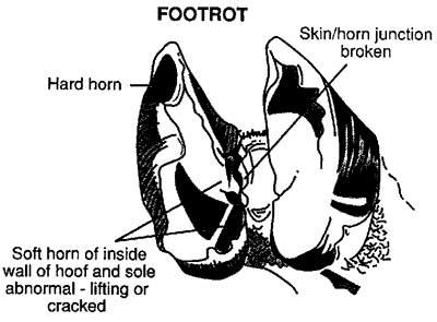
    Clinical Signs:
    Animals infected with footrot can be mildly to severely lame in one or more feet. The area between the toes is inflamed and moist. The problem spreads to the sole of the hoof causing it to separate from the underlying tissues. A characteristic foul odor and some discharge are often noticed.

    Diagnosis:
    Cases of footrot are often diagnosed based on clinical signs and evidence of damage to the hoof. Footrot tends to infect more animals in the flock/herd at one time than does footscald. Cultures of samples taken from problem areas can be used to identify B. nodosus. Problems such as laminitis, bluetongue, and foot-and-mouth disease may also have symptoms similar to footrot.


    Treatment:
    Aggressively trim the feet of every animal in the flock/herd. Lame animals or animals with obvious signs of footrot should have any problem areas of hard and soft hoof completely removed. This is probably the most essential part of the treatment program. Trimming removes cracks and areas for the bacteria to hide in and also allows medications to reach the bacteria. When small numbers of animals are infected, sprays and brushes can be used to apply topical medications such as antibiotics (tetracycline) or antiseptics (copper sulfate, zinc sulfate, or 5% formalin). Remember to disinfect all hoof trimming equipment after trimming each, individual animal’s feet to prevent the potential spread of infection.

    After completely trimming all animals (infected and non-infected), soak all hooves for 5 minutes in a foot bath containing a 5% formalin, 10% zinc sulfate, or 10% copper sulfate solution. Zinc sulfate is as effective as formalin, is safer to use, and less irritating. Because it can stain the wool or hair, copper sulfate should be used with caution. Separate infected and non-infected animals and repeat the treatment on both groups of animals one week later. Infected animals should receive additional footbaths at 7-day intervals for several more treatments. Severely affected animals may need to be placed in a foot bath for up to an hour at a time. Turn apparently cured sheep/goats into an uncontaminated area, and re-examine all sheep/goats on a regular basis. Regular examination is essential because some sheep/goats thought to be clean may actually still be infected. With time and moist conditions, these animals may reinfect other sheep/goats. In problem flocks and herds, force sheep/goats to move through a 10% zinc sulfate solution daily for 30 days. This has become a very successful treatment. Sell any animals that are persistently lame. If all sheep/goats are sold, wait 3 weeks before bringing in new sheep/goats. Treatments using injectable antibiotics can also be used. Penicillin and long-acting oxytetracycline are commonly used.

    * No one method of treatment seems to be completely affective. This means that a combination of trimming feet, footbaths, and vaccinations should be part of every treatment program.

    Prevention:
    In flocks/herds that are footrot free, maintaining a closed flock/herd is essential. If this is not possible, all new animals that will be potentially added to the flock/herd should be examined for evidence of foot problems. If an animal is suspected of having footrot, it should be isolated for at least 2-3 weeks while being treated with footbaths or injectable antibiotics. Never use equipment (tracks, hoof trimming tools, etc.) that could have been contaminated by infected animals.

    Proper feet trimming, keeping animals out of wet marshy areas, regular footbaths during the wet seasons, and routine culling of lame animals are all essential prevention ideas. Vaccines are also available that can be used to prevent footrot. The two products currently on the market are Volar (page G926) and Footvax (page G244). The Volar product can be used to prevent infections of Fusobacterium necrophorum. Footvax is used to prevent infections of Bacteroides nodosus. Some animals may develop injection site lesions due to these vaccines; this should be considered when administering them to show animals.
     
  2. Footscald (interdigital dermatitis):
    Introduction/Causative Agent:
    The term footscald can be used to indicate irritation found between the claws. Many times footscald is caused by the organism Fusobacterium necrophorum. This bacteria is found throughout the environment where sheep and goats are found. All animals are exposed to the organism, and most do not develop problems. However, wet muddy environments and possible injury to the foot allow the right situation for the organisms to infect the foot. Some reports indicate that the organism can remain in the soil for years, that the organism is present in almost all sheep and goat environments, and that it is virtually impossible to remove. Some researchers also consider infections of the non-proteolytic (non-tissue destructive) strains of Bacteroides nodosus to be "scald." Infections with F. necrophorum can lead to problems with footrot.

    Clinical Signs:
    Most animals with footscald are only mildly lame. Footscald (unlike footrot) involves only the tissues between the claws and not the claw itself. The problem usually goes away once the animals’ environment becomes drier.

    Treatment/Prevention:
    Most of the treatment and prevention ideas discussed for footrot also apply for footscald.
     
  3. Abscesses and Ulcers: These are often discovered when inspecting the foot or using hoof testers and appear as pockets of infected material with abnormal coloration of surrounding hoof tissue. Often, these are associated with rough surfaces, nails, excessive toe growth, poor conformation, and problems with laminitis. Treatment for most abscesses involves cleaning/paring out the abscess to promote drainage and antibiotic treatment. Placing the unaffected claw on a block to prevent weight bearing on the affected claw is also used to promote healing.
     
  4. Fractures: The clinical signs include mild to severe lameness and swelling. In some cases, the animal may refuse to bear weight on the limb. Treatment depends on the location of the fracture and should involve a veterinarian.
     
  5. Laminitis - This can be a very common problem on a flock/herd-wide basis. The cause of laminitis on a flock/herdwide basis is usually a result of a diet high in carbohydrates and low in fiber. This causes a decrease in rumen pH and a potential laminitis problem. Laminitis on an individual animal basis can be caused by mastitis, rumenitis, grain overload, pneumonia, and retained placenta. Clinical signs include shifting leg lameness, tenderness, pink areas around the white line, and abnormal rings of hoof growth below the coronary band. Treatment involves the administration of phenylbutazone (bute) and correcting the diet problems or individual animal diseases.