Introduction:
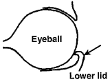
Entropion is found in all goat and sheep breeds. The condition is non-lethal; however, entropion can be passed on genetically and any animal with this problem should not be retained for breeding purposes. Some genetic family lines of whiteface breeds of sheep tend to be more prone to this fault. Some people also think that this problem can be caused by environmental conditions such as the extended use of heat lamps or ultraviolet irradiation.Clinical Signs: Entropion is a situation where the eyelid(s) are turned in (inversion), causing irritation and abrasions to the surface (cornea) of the eye. This causes severe tearing (lacrimation) and usually occurs in young animals within the first week to 1-2 weeks of life. Generally, the most common location of this condition is found on the lower eyelid. Either one (unilateral entropion) or both (bilateral entropion) eyes may be involved with this condition. If uncorrected, the inverted eyelids damage the cornea of the eye and cause ulceration, cloudiness of the cornea, and sometimes blindness.
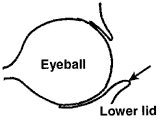
Ectropion is another, less common, eyelid problem. This is a condition where the lower (and sometimes the upper) eyelid is turned outward and becomes a pouch or pocket for debris that can also damage the cornea and eye.
 |
 |
| Entropion | Ectropion |
Diagnosis:
Diagnosis is made by closely examining the eyelids of all animals at docking, castration, or tagging time. Any animals that have tearing (lacrimation) that soils the hair on the side of the face should be closely examined. If there is any evidence of the lashes of the eyelids contacting the surface of the eye, the animal has entropion.Treatment:
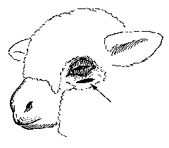
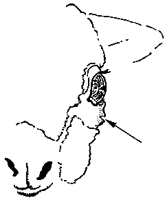
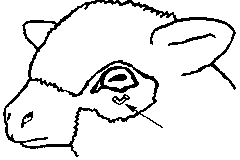
In some minor cases of entropion, correction can be made by injecting 1-2 mLs of a long-acting, slowly absorbed antibiotic (procaine penicillin) under the skin of the affected lid. This will help distend and roll the eyelid outward into a proper position and help alleviate some of the irritation. Sometimes staples, suture, or clips are applied to the skin surface of the problem eyelid. In these cases, the tension of the clip or staple alone may pull the lid into proper placement. In many of the younger animals, these procedures are often all that is needed to correct the problem.In more complicated cases, this condition is corrected by surgically removing an elliptical portion of the skin under or over the problem eyelid (see figure 1) with a knife or curved tip surgical scissors. The wound is sometimes left open to heal and antibiotics are applied. As the wound heals, the increased tension pulls the lid into proper placement. Stainless steel surgery clips (see figure 3), staples, or stitches (suture) are also utilized to close the created skin defect and help correct the inversion.
These procedures are most effective if performed early in the life of the newborn, especially while the skin is softer and more pliable. Advanced cases that require surgery or cases where the eye is damaged, will need antibiotic eye ointment applied 3-4 times a day.
 |
|
 |
|
 |
|