This is a view of the equine uterus from above, and behind.
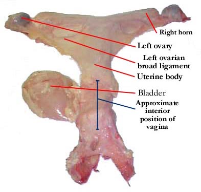
This is a view of the equine uterus from above, and behind.
This is a view of the same uterus, from below. Here the urethral connection of
the bladder can be seen clearly (the bladder is again displaced to the left),
and the body and horns of the uterus are more clearly differentiated from the
broad ligament.
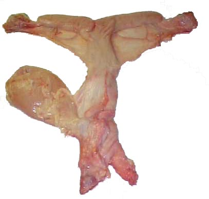
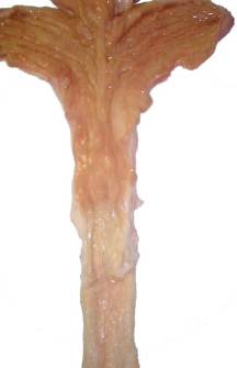
In this image, the bladder, fat and broad ligament have been stripped away, clearly revealing the uterine body and horns.
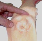 |
On the left, the cervix is clearly exposed, and can be seen as the raised section between the forefinger and thumb of the hand. |
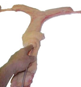 |
In this image, the insemination pipette is seen being introduced through the cervix from the vagina, although the vagina itself has been removed. Note the index finger is introduced slightly (up to almost the first knuckle), and is being used to guide the pipette into and through the cervix, as it would when carrying out an insemination. |
 |
On the left can be seen the different textures of the uterine/cervical/vaginal tissue. The darker section at the top is the endometrium, the light section in the middle of the image is the cervix, and the lower section is the vagina. |
| The ovary (seen on the right) is roughly kidney-shaped, with the depression in the center of the lower side being the ovulation fossa, where the ova evacuated from the follicle (which "implodes" rather than "explodes" upon ovulation), exits on it's way to the uterus after ovulation. The follicle shown here outlined in red is approximately 2 cm, the size being measured from the outer surface to the innermost extent (in this image therefore, from left to right). As a result, ultrasound is the only truly accurate method for establishing follicular size. Determination of size by rectal palpation relies on the assumption that the follicle is spherical in shape, and although this is often the case, it is not always so. Even if the follicle is spherical, it is difficult to determine the exact edge when using palpation. On the other hand, rectal palpation is once again becoming more widely used to determine how close to ovulation the mare is, as the consistency of the follicle softens dramatically close to ovulation, and this can only be established upon palpation. Ultrasound may detect either a "tear-shaping"; or a thickening of the outer membrane of the follicle, both signs of impending ovulation, but these signs are not always present. | 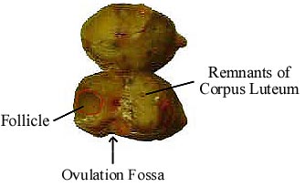 |
With thanks to Canada's Equine Research Center for providing us with the reproductive tract for dissection.
© 1999 Jos Mottershead. May be used upon notification, and with accreditation.