F155
Digestive Tract Problems


megaesophagus | esophagitis | gastritis | ulcers | digestive tract foreign bodies | gastric dilation and volvulus (gdv) | motion sickness | obstructions | inflammatory bowel disease | megacolon | gastrointestinal (gi) parasitism | viral diseases | bacterial diseases


Introduction: The digestive tract of an animal basically begins with the mouth and ends with the large colon. All of the structures between these two points play an essential role in the consumption, breakdown, and utilization of food. Dogs, like horses and humans, are considered simple stomached animals, meaning that their stomachs have only one major compartment. When all the structures in the dog’s digestive tract are functioning properly, the dog is able to gather the necessary nutrients to sustain life; however, problems with anything from the teeth to the intestines can cause weight loss, diarrhea, and vomiting, plus many other signs of problems. The following information will identify some of the more common problems associated with the digestive tract of a dog. The information will start with the esophagus and move through the various parts of the digestive tract.

Esophagus

Introduction: The esophagus is the muscular tube which connects the mouth to the stomach. In people, the tube is oriented up and down when eating, allowing gravity to help direct food into the stomach. In dogs, however, the esophagus is oriented horizontally, and gravity cannot normally play as large a role in its function as it can in people. The muscular action in the esophagus is known as "peristalsis." Peristalsis is a wave of muscular contraction that moves in a front-to-back direction, propelling food into the stomach.

  1. Megaesophagus: Megaesophagus is a medical term that means "large esophagus." Megaesophagus occurs when the tube’s muscle wall loses the ability to move food down into the stomach and stretches out into a much larger size than normal. Megaesophagus occurs as three different types: congenital, acquired idiopathic, and secondary.

    Clinical Signs
    : The most common clinical sign associated with megaesophagus is regurgitation. Regurgitation is different from vomiting in several very important ways. Vomiting occurs when the body expels stomach contents and occurs as a deliberate action. Warning signs of nausea and abdominal heaving are usually seen or felt. Regurgitation occurs when food has been sitting in the esophagus, unable to arrive at the stomach. When the esophagus expels food, it usually occurs as an almost passive action with little to no warning at all. The animal opens its mouth and suddenly expels whatever it has eaten. Regurgitated food is usually undigested and may even have a cylinder-like shape. Regurgitation can occur minutes to hours after eating.

    Aspiration pneumonia (a specific type of pneumonia that results when the animal inhales its own food/water) is very common with megaesophagus. Aspiration pneumonia is very similar to other types of pneumonia, accompanied by shortness of breath, coughing, and fever.

    Dogs with megaesophagus may or may not be able to pass enough food into the stomach and intestines to survive. Dogs with only mild cases may show no signs at all of malnourishment. On the other hand, dogs with severe cases may become malnourished or even starve to death if little to no food passes from the esophagus into the stomach. Because the body tries to obtain the nutrition that it needs, these dogs may have a tremendous appetite.
     
    1. Congenital Megaesophagus is first seen in young puppies, usually between 6 and 10 weeks of age. Breeds affected by congenital megaesophagus include miniature schnauzers, fox terriers, Great Danes, German shepherds, Labrador retrievers, and Irish setters. Many puppies do recover on their own as they mature. While spontaneous recovery can occur up to 12 months of age, if a puppy with congenital megaesophagus has not significantly improved by the time it is 6 months old, the outcome is usually poor.
       
    2. Acquired Idiopathic Megaesophagus is probably the most common type of megaesophagus seen in dogs. This type occurs with no known underlying cause, and generally affects large-breed dogs between 8 and 14 years of age. Treatment of this type of megaesophagus focuses on supportive care only, because there is currently no known way of correcting the disease. Prognosis is often poor.
       
    3. Secondary Megaesophagus describes any megaesophagus where another disease process is causing the problem. There is a long list of possible conditions and diseases that have been known to cause a secondary megaesophagus. Finding the exact underlying cause may be extremely time-consuming and expensive. Diseases and conditions such as systemic lupus erythematosus, distemper, myasthenia gravis, hypothyroidism, Addison’s disease, gastric dilatation/volvulus (GDV), lead poisoning, and many others have been known to cause secondary megaesophagus. Any breed or age of dog may be affected, and treatment focuses on the underlying cause as well as supportive care. The outcome and prognosis of dogs affected by secondary megaesophagus vary greatly depending on the cause. Sometimes the condition can be controlled or even resolved, while in other cases the problem may persist in spite of proper treatment.

      The most common underlying disease causing secondary megaesophagus is a condition called myasthenia gravis. Because this is a relatively common underlying cause of megaesophagus, some description of this disease will be made at the end of the megaesophagus section.


    Diagnosis
    : Diagnostic tests used for megaesophagus include physical examination, CBC, serum chemistry profile, urinalysis, and chest x-ray films (often with contrast dye mixed with a meal to highlight the esophagus). Most of the time megaesophagus can be seen on the chest films. More specific diagnostic tests may then be performed in the search for an underlying cause. This portion of the diagnostic workup is important because sometimes if an underlying cause can be found, the outcome may be significantly better. Endoscopy (sometimes with biopsy of the inner lining of the esophagus), AChR antibody test for myasthenia gravis, thyroid hormone levels, and an ACTH stimulation test for Addison’s disease are other tests that should be strongly considered in the patient’s diagnostic plan.

    Treatment
    : Megaesophagus treatment usually focuses on the underlying cause, if any, and on supportive care. Dietary therapy is a key part of supportive care for megaesophagus. Small, frequent feedings of a high-calorie diet, sometimes prepared as a partially-liquid gruel and fed with the dog in an elevated position, can be very helpful in preventing malnourishment. Many dogs can learn very quickly to eat while standing with the front paws on the edge of a counter or table top. This allows gravity to help pass the food into the stomach. Some patients may require a feeding tube surgically placed in the digestive tract in order to survive. Aspiration pneumonia is treated with antibiotics, rest, and humidification. Attempts at treatment with other types of medication and surgery have been tried, but most often without significant improvement.

    Prognosis of megaesophagus depends on the type and underlying cause. At the time of diagnosis, this condition usually warrants a guarded prognosis, but may improve with time; this is the case with some congenital megaesophagus puppies that outgrow their disease. Others will worsen in spite of all possible treatment.
     

    1. Myasthenia gravis is a complex disease in which nerves lose the ability to stimulate muscle tissue. This occurs because of the destruction of important molecules at nerve-muscle junctions by the dog’s own immune system. The esophagus muscle tissue seems to be especially prone to the effects of this disease. As the lack of nerve communication increases, the esophagus relaxes and "balloons" out, becoming a large distended chamber. This disease can occur in adult dogs of all sizes and ages, but especially in German shepherds and retrievers. The condition is more likely to occur in dogs that are either less than 5 years of age or over 8 years of age. Many dogs develop muscle weakness throughout the body, while others may only develop a weak esophagus.

      Diagnosis: The most common test used to diagnose myasthenia gravis is a serum test that measures the antibodies directed against the communication molecules (acetylcholine receptors). The test is called acetylcholine receptor (AChR) antibody radioimmunoassay.

      Treatment: Treatment of myasthenia gravis consists of medications that help increase the ability of the nerves to stimulate the muscles of the body. Pyridostimine bromide (Mestinon) is a drug that has been helpful in many cases. Corticosteroids such as prednisone are also used in an attempt to suppress the immune system’s attack on the important acetylcholine receptors. Sometimes myasthenia gravis will resolve on its own, while other affected animals will stop responding to any treatment at all and progressively deteriorate. Response to treatment is variable, and prognosis is always somewhat guarded when this disease is diagnosed.
       
  2. Esophagitis is the medical term for inflammation of the esophagus. This condition usually occurs when the dog swallows a substance or object that is irritating to the inside lining of the esophagus. Cleaning agents that contain acids or bases, other corrosive products, sharp or oversized objects, and even some prescription drugs are sometimes eaten and swallowed by dogs and can cause esophagitis to some degree. Doxycycline is an antibiotic (related to tetracycline) that has especially been known to cause esophagitis in animals when not swallowed all the way into the stomach. The esophagus does have wonderful capabilities to withstand trauma, and most minor injuries heal quickly with no obvious clinical signs.

    Clinical Signs
    : Problems associated with esophagitis include regurgitation, excessive saliva production (saliva may contain blood and often has a thick, stringy appearance), difficulty swallowing, and decreased appetite. If the inner lining of the esophagus becomes severely inflamed and begins to form ulcers, there may be some obvious pain when the animal’s neck is felt from side to side just above the airway. Scar tissue may form from severe esophagitis and result in strictures (narrowing of the esophagus with scar tissue). Esophageal strictures are difficult to treat and may cause severe swallowing problems for the animal.

    Diagnosis
    : It is much easier to diagnose esophagitis when it is known that the animal swallowed something such as a household cleaning agent. Treatment can be started immediately without waiting for any further tests. Physical examination and x-ray films, sometimes with a dye/food mixture in the esophagus, can be helpful in some severe cases. Endoscopy, sometimes with sampling of the inner lining of the esophagus, is the best way to accurately diagnose esophagitis.

    Treatment
    : Treatment of esophagitis depends upon the case. Some patients with mild esophagitis may not require any treatment at all. If a corrosive or irritating material has been swallowed, such as an acid, it is important not to make the animal vomit. Bringing the damaging substance back up again only causes additional injury as it contacts the esophagus twice instead of once. A poison control center may have helpful advice if the name or active ingredient of the damaging substance is known. Diluted vinegar can be given to counteract basic substances, milk of magnesia for acidic substances, and activated charcoal can be given to bind poisonous substances so that they cannot be absorbed into the body. A veterinarian may need to pump the animal’s stomach in order to remove the dangerous substance. Resting the esophagus by withholding food and water for several hours following any swallowing injury is helpful when starting the healing process. In some severe cases, a feeding tube surgically placed in the stomach may be necessary for a period of time to give prolonged rest to the esophagus. Sucralfate (see page H792) is a specific medication that can greatly help the healing of the esophagus. Prednisone or other corticosteroids may be prescribed to help prevent the formation of scar tissue strictures after severe injury to the esophagus.


Stomach and Intestines

  1. Gastritis: Acute gastritis/gastroenteritis means "sudden inflammation of the stomach and/or intestines." This is an extremely common occurrence in small pets and is probably the most common cause of abrupt vomiting in dogs. The cause of acute gastritis or gastroenteritis is usually the reaction in the digestive tract to something the dog has eaten. The ingestion of spoiled food, medications, toys, plants, and fungi (mushrooms, toadstools) can cause gastroenteritis. Allergies or reactions to certain substances in food such as corn or wheat are also common ways that a dog can develop gastroenteritis.

    Clinical Signs
    : The number one clinical sign seen with acute gastroenteritis is vomiting. Occasionally, the dog may simply be listless and refuse to eat without vomiting, but this is rather uncommon. The vomit may contain food and often has a yellow or green tinged color, frequently with white foamy or frothy matter. Small amounts of blood may also be present. Most dogs do not feel well and may walk with a hunched back, cry when the abdomen is touched or pressed, and do not want to move or eat as much as they normally would. Diarrhea is seen with some cases of gastroenteritis, but is not as common as vomiting. Development of a bleeding ulcer in the stomach or intestines can occur and may lead to a dark discoloration of the stool known as melena (passage of digested blood). Acute gastroenteritis usually lasts between 1 to 5 days in dogs.
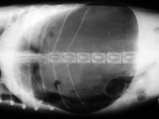
    Diagnosis
    : Acute gastroenteritis is best diagnosed by a veterinarian who can perform a thorough physical examination and other tests if necessary. Many diseases of the digestive tract which cause the same symptoms as simple gastroenteritis are much more serious and even life-threatening. A veterinarian can usually distinguish between the less severe and more severe situations with a physical examination and other tests. X-ray films of the stomach and intestines can be extremely helpful in determining the seriousness of the situation. X-rays are usually normal with acute gastroenteritis. Bloodwork (CBC and serum chemistry panel) and urinalysis are other very important and helpful tests that can point to other affected systems such as the liver and/or pancreas, and can help the veterinarian know how aggressively to treat the animal. Other tests that can be performed are endoscopy of the digestive tract and ultrasound of the abdomen.

    Treatment
    : Treatment of acute gastroenteritis depends on current condition of the animal. Prolonged vomiting and diarrhea can quickly lead to dehydration. Old, small, or dehydrated patients may require more aggressive treatment than others. Fluid replacement therapy should be addressed in all patients with acute gastroenteritis. Oral fluids may be sufficient in mild cases and can be administered by frequently offering small amounts of ice. Fluids should be given under the skin or intravenously in patients that are more severely ill or dehydrated. Potassium is lost through vomiting and many patients with acute gastroenteritis are low in body potassium levels. Potassium is measured on routine bloodwork and can be replaced either orally or in replacement fluids. Diet is a second very important part of treating acute gastroenteritis. It is usually best to withhold food until the patient has stopped vomiting for at least 12 hours. When food is offered again, low-fat foods rich in carbohydrates are recommended. Cooked rice or pasta and low-fat cottage cheese offered in small, frequent quantities have been very beneficial in the recovery stages. Antiemetic therapy (medication that stops vomiting) is not usually recommended in patients with acute gastroenteritis except in very special cases. Generally, any dog that requires use of these medications is best kept hospitalized under the direct care of a veterinarian. Because ulcers of the stomach and/or intestines may result in some cases, antibiotics are often used in the treatment of acute gastroenteritis. Antacids and pain killers can also be used, but non-steroidal anti-inflammatory drugs (NSAIDs) such as aspirin or carprofen should be used with caution because they can cause ulcers to form in the digestive tract.

    Prognosis for acute gastroenteritis is excellent in most cases. While some animals can be treated at home for this condition, others will require hospitalization and direct care of a veterinarian. All animals with symptoms of abdominal pain and vomiting should initially be examined by a veterinarian to help ensure that another more serious condition does not exist.
     
  2. Canine hemorrhagic gastroenteritis (HGE) is a sometimes fatal disease that occurs in dogs. While any dog breed can develop HGE, small breed dogs are at higher risk, especially schnauzers, poodles, and dachshunds. The cause of HGE is not as yet known, but there is speculation that Clostridium perfringens bacteria may be at least partially to blame.

    Clinical Signs
    : Hemorrhagic gastroenteritis is a disease where the inner lining of the intestines dies and sloughs off rather suddenly, leading to vomiting and diarrhea. The vomit and especially the stool that are expelled usually contain large amounts of blood. The diarrhea especially has been accurately described as having a "raspberry jam" appearance. The dog becomes very depressed and weak in a very short period of time. Much of the diarrhea contains sloughed intestinal lining and carries an unusually foul, "dead" smelling odor. Without treatment, the dog will usually go into shock within hours. Many, if not most dogs will die from HGE without proper and immediate therapy. However, the expected outcome is usually good if the patient is treated rapidly and aggressively.

    Diagnosis
    : There is no single, specific test that is used to diagnose canine hemorrhagic gastroenteritis. A dog with a history of sudden bloody diarrhea and vomiting, especially if it is a small breed, should be suspected of having HGE. A physical examination should be performed by a veterinarian as soon as possible (using an emergency service is recommended if the problem occurs after-hours). Bloodwork is helpful in making a diagnosis of HGE, especially a packed-cell volume (see page D120). The packed-cell volume in these cases is usually abnormally high. Cytology and culture of the bloody stools can also be helpful, especially if Clostridium perfringens bacteria are found in high numbers. X-ray films of the abdomen may be performed to look for other causes of bloody diarrhea and vomiting, but are usually normal in animals with HGE. Parvo testing should also be performed, especially in young dogs that have not been previously vaccinated and are experiencing bloody diarrhea and vomiting.

    Treatment
    : Very aggressive intravenous fluid therapy is the primary treatment for HGE. The packed-cell volume is usually monitored very closely during treatment and can be used as an effective gauge for how fast the intravenous fluids need to be given. Antibiotics are also an important part of treatment. Occasionally, if enough blood is lost through the digestive tract, a blood transfusion may be needed. The disease generally resolves within 24 to 48 hours, although the patient may take several more days to return to a normal state.
     
  3. Ulcers: Ulcers of the stomach and intestines are relatively common in dogs and affect animals in much the same way they affect people. Consuming certain drugs is probably the most common reason for ulcers to develop in the digestive tracts of dogs. Aspirin, ibuprofen, acetaminophen (Tylenol), and other over-the-counter pain killers are perhaps the most notorious drugs for causing ulcers in the digestive tract. These drugs are known together as "non-steroidal anti-inflammatory agents" (NSAIDS) and are discussed in Orthopedic Problems F610. Veterinary prescription NSAIDS such as carprofen (Rimadyl), etodolac (EtoGesic), and deracoxib (Deramaxx) are less likely to cause digestive tract problems than the over-the-counter human products, but can lead to vomiting and diarrhea, sometimes with digestive tract ulceration. Other causes of stomach/intestine ulcers in dogs include stress, infections, eating rotten or non-food items, some types of cancer (especially mast cell tumors), liver failure, kidney disease, and disorders of the neurologic system.

    Clinical Signs/Diagnosis
    : The most common clinical sign of ulceration of the stomach in dogs is vomiting. Decreased appetite, abdominal pain, listlessness, melena (dark, tarry stools), and blood in the vomit are also commonly observed. Bleeding from a digestive tract ulcer can be significant, occasionally leading to death. Diagnosis of digestive tract ulceration is based on the patient’s history, physical examination, bloodwork, x-ray films (preferably with barium contrast dye in the stomach and intestines to outline the shape of the inner lining of the digestive system), and sometimes endoscopy.

    Treatment
    : Treatment of digestive tract ulcers depends on the cause of the problem and the severity of the ulcers. Removing the source and cause of the ulceration is important to a successful treatment program. Medical therapy for stomach ulcers is usually successful. Antacids such as aluminum hydroxide, magnesium hydroxide, ranitidine (Zantac - see page H993), cimetidine (Tagamet - see page H800), famotidine, and omeprazole are often central to the treatment of stomach ulceration. The acid in the stomach constantly irritates the damaged portion of the inner lining of the stomach, making it very difficult for the ulcer to heal itself. Antacids lessen this irritation by either neutralizing the stomach acid or decreasing its production. Another commonly used drug in the treatment of stomach ulcers is sucralfate (Carafate). Sucralfate (see page H792), which must be taken orally, sticks tightly to ulcerated areas in the stomach, acting as a protective "band-aid." It has even been used at higher doses for bleeding ulcers to prevent hemorrhage. Misoprostol is a drug that is used more in the prevention of NSAID-caused stomach ulcers than for treatment. Misoprostol increases the flow of blood to the inner lining of the stomach and slows the secretion of acid. It is often given with an NSAID to help protect the patient against stomach ulcer formation.

    Antibiotics are often given for secondary infection of an ulcerated portion of the digestive tract. Blood transfusions are occasionally required to treat a patient with severe blood loss from a bleeding ulcer. Ulcers can be removed surgically if necessary to save the patient from severe bleeding or if the ulcer threatens to tear completely through the stomach, both of which are life-threatening situations.
     
  4. Digestive tract foreign bodies are very common in dogs, especially in puppies and large-breed dogs. Some dogs will chew up and eat almost anything, including toys, glassware, clothing items, shoes, string, furniture, electrical equipment, books, rocks, and other non-food items. Any non-food item that is found in the digestive tract is called a GI (gastrointestinal) foreign body. Much of the time, foreign bodies can pass on their own without any major intervention. In fact, many dog owners do not even realize the family pet has eaten something it shouldn’t until a missing toy or other item turns up in the pet’s morning bowel movement. If a foreign body becomes lodged or blocks a portion of the digestive tract, however, the situation may rapidly become fatal if not treated.

    Clinical Signs/Diagnosis
    : Signs of a digestive tract foreign body include vomiting, diarrhea, abdominal pain, decreased appetite, fever, listlessness, shock, and even death. Diagnosis is based on history, clinical signs, physical examination, bloodwork, abdominal x-rays, ultrasound, and exploratory surgery.

    Treatment
    : Depending on the item ingested, location within the body, and degree of illness treatment varies. Dehydration is corrected with fluids and electrolytes if needed, and antibiotics are usually given for treatment of secondary infections in the digestive tract. Surgery is performed early on if a blocked bowel is noted on x-ray films or ultrasound, or if the item ingested will obviously not pass on its own. Items that have made their way into the large intestine and colon are usually left alone to pass on their own. Linear foreign bodies such as string, shoelaces, or shredded towels are particularly damaging to the digestive tract because they can lead to "accordion" formation or bunching up of the intestine along the foreign object, and very large sections of intestine can die in this manner. If the patient can be kept stable and the ingested item is not causing blockage of bowel and appears to be able to pass on its own, a waiting period may be a sufficient treatment plan. X-ray films should be taken to monitor the progress of the foreign body. If there has been no progress made after 48-72 hours, surgery should be scheduled. When surgically removing a GI foreign body, the entire digestive tract is carefully examined. If death of a portion of the digestive tract has occurred, it must be removed. Removal of sections of the digestive tract is very challenging and time-consuming. Any digestive tract surgery is generally fairly expensive and the recovery period can be lengthy.
     
  5. Gastric dilatation and volvulus (GDV) is a serious condition in dogs that can lead to shock and death if not promptly treated. GDV occurs when gas rapidly accumulates in the stomach causing the stomach to twist and rotate out of its normal position. The blood vessels of the twisted stomach can become pinched, disrupting normal blood flow and producing the effects of shock and even death.

    The precise cause of GDV remains unknown. It is generally thought that the build-up of air in the stomach is caused by the dog swallowing air while eating, followed by the inability to release the air through eructation (or belching). However, how this accumulation of air leads to the twisting of the stomach is unknown. Although scientific research has been unable to pin-point the cause of GDV, several risk factors have been found to increase the probability of developing GDV. These risk factors include the following:
     
    • Breed type - GDV is usually manifested in dogs that have a deep and narrow chest, such as the Great Dane, Irish Wolfhound, Standard Poodle, Irish Setter, German shepherd, etc. However, many types of breeds can develop GDV.
    • The occurrence of GDV in a relative - If the dog has a close relative (such as a mother, father or sibling) that has a history of GDV, the probability of developing this condition greatly increases.
    • Eating rapidly
    • Having a diet of mostly dry food - Both eating rapidly and consuming a primarily dry-food diet appear to increase the volume of air taken into the dog’s stomach.
    • Having a nervous or fearful disposition - Studies have shown that dogs with a fearful and/or nervous personality are more likely to develop GDV than dogs with a generally happy, calm disposition.
       
    Clinical Signs: The accumulation of gas in the stomach will usually produce visible swelling of the abdomen that feels tense to the touch. The dog may also show signs of discomfort and distress (such as whining). The dog may become restless, unable to find a comfortable position, and may attempt to vomit repeatedly without producing anything. Collapse and death may occur within hours to days after developing GDV.

    Treatment
    : If the dog is suspected of having GDV, he/she should be taken to the veterinarian immediately for treatment. Initial treatment for GDV often includes passing a stomach tube or inserting a needle into the dog’s abdomen to release the gas build-up. Surgery is generally needed to untwist the stomach and return it to its normal position. The veterinarian can also perform a gastropexy. This is a procedure in which the stomach wall is surgically attached to the abdominal wall in an effort to help prevent the stomach from twisting out of position in the future.

    Some suggestions that might help reduce the probability of developing this condition include the following:
    • Slow the dog’s eating speed.
    • Feed the dog small meals.
    • Avoid excessive water intake immediately before or after feeding.
    • Avoid strenuous exercise immediately before or after feeding.
       
    Dog owners should watch for signs of GDV, especially if their dog has a deep, narrow chest and/or relatives with a history of GDV. Recognizing and responding promptly and properly to the onset of GDV may save a dog’s life.
     
    This is an x-ray of a dog with a GDV. The dark, round area is the distended stomach.


  6. Motion sickness is extremely common in dogs. Vomiting, excessive salivation, yawning, uneasiness, and crying in a car, boat or aircraft are the most commonly observed signs. The motion stimulates sensors in the inner ear which has connections to the parts of the brain that control nausea. Animals may become afraid of a car or other vehicle after repeated episodes of motion sickness, and may even become sick before the vehicle starts moving. Medications that are commonly used for carsickness in dogs include diphenhydramine hydrochloride (Benadryl) at 25 mg for small dogs and 50 mg for medium to large dogs given 1 hour prior to traveling, and acepromazine maleate. Acepromazine is a prescription drug that must be obtained through a veterinarian (see page H10).


Intestines and Colon

  1. Obstructions: Intestinal obstruction and surgical diseases of the intestines are usually life-threatening emergencies. Obstruction of an intestine causes gas and food contents of the digestive tract to back up, swelling the intestine and causing severe pain. Bacteria and other organisms normally live in the intestine and help in the breakdown of food. The intestinal wall is normally a strong effective barrier that prevents these bacteria from entering into the rest of the body. When obstructions or other severe diseases of the intestines occur, however, the intestinal wall becomes inflamed, stretched, and even torn, allowing bacteria to cross into the body. Severely stretched, inflamed, and dying intestines are not able to prevent bacteria from crossing deep into the walls and getting into the veins that drain blood from the digestive tract. These veins go directly to the liver for filtering. Large numbers of bacteria flowing into the liver can overwhelm the organ’s filtering ability and pass into the rest of the body. Bacteria floating around the bloodstream can then trigger a violent reaction by the body’s immune system. This set of conditions is called septicemia and septic shock. An animal in septic shock is in critical condition and will usually die within 24 hours without aggressive medical intervention.

    If an intestine becomes torn by a sharp object or if the gas swelling stretches it to the point of rupture, the bacteria-filled fluid inside spills into the abdomen. Severe infection then results in the abdominal cavity, leading to a condition called peritonitis. Septicemia and septic shock quickly develop from peritonitis. For almost any type of obstruction or swelling of the intestines, surgery is needed to correct the problem. The animal should first be stabilized and brought out of shock if needed. Then surgery must be performed to remove the source of obstruction and any dead or dying portions of the digestive tract. This type of surgery is often very challenging, time-consuming, and expensive. Following are a few varieties of intestinal obstruction seen in canine patients.
     
    1. Foreign objects - Non-food items that the dog has eaten are perhaps the most common cause of intestinal obstruction in dogs, especially in puppies. Some dogs will eat almost anything, and care must be taken by a dog’s owners to keep the pet’s living environment clear of dangerous objects that may be chewed on and swallowed. Bones are a rather common foreign object that may become stuck in the intestines and should be avoided. Children’s toys, clothing items, bedding, sticks, rocks, food wrappings, hairballs, fruit pits, decorations, books, furniture, and fish hooks are just some of the objects that have been retrieved from the digestive tract of dogs.
       
    2. Linear foreign objects - Linear foreign objects are a special category of foreign objects swallowed by dogs. Cats are more likely to eat string or other long objects than dogs are, but linear foreign objects are commonly seen in both species. A string-like foreign object will usually catch on some portion of the digestive tract and dangle itself through the intestines. As the intestines go through their normal movements trying to pass the object, they catch in various places along the linear foreign object and bunch up along its length, much like an accordion. As the intestines become inflamed and swell, a great deal of tension is put upon the intestines where it contacts the linear foreign object and the object begins to cut through the intestines. Surgically, this is one of the most challenging types of obstruction to repair, and the animal may end up losing very large portions of their digestive tract.
       
    3. Intussusception - Intussusception is the medical term for "telescoping" of the intestines. This condition is most common in puppies less than one year of age. The length of the intestine rolls itself into the intestine on either side of it, forcing the receiving side to swell. As the intestines become more inflamed and swollen, the blood supply to the kinked portion of the intussusception is cut off and the bowel begins to die. Sometimes the surgeon can unroll the bowel and save the affected portion, but quite often the entire section must be removed.
       
    4. Twisted bowel (volvulus) - Twisting of a section of bowel can occur, but is uncommon as a sole problem. It is usually accompanied by other types of digestive tract disease. Twisted bowel can die very quickly and septicemia and septic shock can take a dog’s life in 24 hours or less from the time that the twisting first occurred.
       
    5. Stricture (scar tissue) - Scar tissue can develop in the intestinal tract from any type of injury or disease, but is most common after surgery on the intestines. Veterinary surgeons are aware of this complication and take precautions when correcting intestinal problems in order to avoid scar tissue blockage down the road. If scar tissue does cause blockage, however, it usually comes on gradually as a partial blockage slowly getting worse and worse with time. The pet will generally go through periodic bouts of illness that may gradually worsen. Sometimes a small foreign object such as a hairball may lodge itself in a portion of the intestines narrowed by scar tissue formation and cause sudden, complete blockage. Most of the time, scar tissue can be removed by surgery, but the increased risk of new scar tissue formation with a second surgery is unavoidable.
       
    6. Abscesses - Occasionally, infections of the intestines will result in abscesses in the walls of the intestines or adjacent structures. Abscesses may swell and block off the normal flow of the intestines, leading to obstruction. Perhaps the most common spot for this to occur is in the dog’s cecum, a small pouch of dead-end bowel at the connection between the small and large intestines. In people, this pouch is somewhat smaller and is called the "appendix." Appendicitis is more common in people than is infection and inflammation of the cecum in dogs. Removal of the inflamed and infected cecum may be necessary to correct obstruction in this area.
       
    7. Hernias - Hernias are defects or tears in the body wall and can occur at the umbilicus ("belly button"), inguinal area (groin), and in the diaphragm. If a defect or tear somewhere in the abdominal cavity is large enough to allow a loop of intestine to pass through, it can become a severe problem. If the intestinal loop twists inside the hernia and becomes entrapped, it will become swollen and obstructed. This most commonly happens in inguinal hernias and tears in the diaphragm. Surgery to repair the hernia as well as to restore health to the digestive tract is the common treatment.
       
    8. Neoplasia - Cancers of the intestines will cause obstruction. Sometimes symptoms of a sudden obstruction are the first signs a pet’s owners may notice. Cancers of the intestines may be either benign or malignant and may arise from the gland tissue in the intestinal walls (adenoma, adenocarcinoma), from the smooth muscle layer (leiomyoma, leiomyosarcoma), or from the lymphatic tissue (lymphosarcoma). Some types of cancer are very aggressive, such as adenocarcinoma, and may invade other organs and near-by structures. Surgery to remove such cancers can be difficult to impossible. Other types of cancer (lymphosarcoma) may occur diffusely throughout the entire digestive tract and are best treated with chemotherapy rather than with surgery. Smooth muscle cancers and benign cancers are probably the least severe types of intestinal cancer and can be removed surgically.

    Clinical Signs: Signs of intestinal obstructive disease are usually acute (come on suddenly), and include vomiting, abdominal pain, abdominal swelling, loss of appetite, diarrhea, dehydration, and shock. Diarrhea and passage of feces does not rule out an obstruction of the intestines.

    Diagnosis/Treatment
    : Diagnosing an obstruction may be accomplished with x-ray films, ultrasound, endoscopy, or exploratory surgery. Treatment almost always will include surgery to repair an obstruction and must be scheduled as soon as the patient is stabilized. The expected outcome depends greatly on how much damage has been done to the body by the obstruction and whether septicemia and septic shock have developed. Septic shock must be treated very aggressively; unfortunately, even with aggressive and proper treatments, some studies have shown a survival rate of less than one in three. Cancers of the intestines carry their own expected outcomes, depending on the type of cancer and whether metastasis and spreading of the cancer has occurred. 
     

  2. Inflammatory bowel disease (IBD) is a term applied to a group of diseases where the walls of the intestines become invaded with white blood cells and are chronically inflamed. IBD is perhaps the most common cause of chronic vomiting and diarrhea in dogs. A variety of causes can allow white blood cells to invade the intestinal walls. Some of these causes include reactions to diet, infections with parasites or bacteria, and intestinal cancers. Portions of the digestive tract may be affected (i.e. upper small intestine, lower small intestine, large intestine), or the entire system may become diseased all at once. Animals of any age can be affected, but the condition is more common in older patients.

    Clinical Signs
    : Problems associated with IBD can differ slightly depending on how much of the digestive tract is affected. Chronic vomiting and lack of energy are the most common signs, but diarrhea with blood and/or mucus may also occur. Excessive gas, bad breath, anorexia, weight loss, and excessive thirst are sometimes seen. As the intestines become inflamed, they become less permeable to nutrients and are less able to absorb food or water. The animal may begin to starve even when it has access to adequate food.

    Diagnosis
    : Inflammatory bowel disease is diagnosed based on the patient’s history, physical examination, bloodwork, radiographs, endoscopy, and ultrasound, but especially by biopsy and histopathology of the affected intestines. The biopsy sample may be obtained by endoscopy or surgery.

    Treatment
    : Removal of the cause of intestinal reaction is important, but sometimes challenging, and can greatly reduce the severity of disease. Cancers should be properly treated when possible, and infections should be eliminated. Dietary treatment for dogs with IBD is considered a top priority since reactions to diet are commonly the cause of this disease. Carbohydrate and protein sources are important to consider in regulating the diet of dogs with IBD. Rice carbohydrates seem to be absorbed better than other cereal carbohydrates. Simple sugars, especially lactose (milk sugar) should be avoided completely. A good quality protein source that is unlikely to cause a reaction should be used, such as lamb, duck, or fish protein. Many dog food companies create varieties of dog foods using lamb and rice, or other combinations of these carbohydrate and protein sources. Drugs that are used in addition to dietary therapy include steroids (prednisone, prednisolone), azathioprine, metronidazole (an antibiotic with some ability to relieve inflammation), and vitamin therapy. Sometimes broad-spectrum antibiotics are used for long periods of time to control inflammation due to overgrowth of bacteria.

    The outcome of inflammatory bowel disease depends upon the case. Dogs with severe cases have a difficult time and may not respond to any type of medication or diet therapy very well. Sometimes death will occur by starvation in spite of all the best efforts to save the dog. Mild to moderate cases have a fairly good chance of living a normal life with treatment, but can experience recurrence of symptoms from time to time.
     
  3. Constipation is the term for infrequent or difficult passage of feces. Dogs seldom experience the same type of condition that people do with true constipation. A dog that is seen straining to defecate is usually not constipated, but will often have some other condition that has caused inflammation of the large intestine. As animals age, passage of feces may slow down through the digestive tract and bowel movements may become less and less frequent. A higher fiber "senior" or "geriatric" type diet can be used to help with the passage of feces in these patients. Mild laxatives for veterinary patients also exist that may be given on a regular basis to help with the passage of bowel movements.
     
  4. Megacolon is an uncommon condition in dogs that can lead to true constipation. The affected colon tends to stretch and does not effectively expel feces as it should. This condition most commonly occurs later in life, but is occasionally seen as a birth defect. Frequent attempts to defecate are typical, with bowel movements ranging from normal to dry and hard. Sometimes the feces may become watery with mucus and/or diarrhea with secondary inflammation of the colon. Diagnosis is made by physical examination, including a digital exam of the colon, and sometimes x-ray films. Laxatives and a special diet are used first in treating megacolon, but are often unsuccessful. Surgery, often requiring a board-certified surgeon to perform the delicate operation, can be performed when medical treatment fails. The outcome is somewhat guarded in all cases of megacolon.


Digestive Tract Infections

  1. Gastrointestinal (GI) Parasitism: Parasites of the digestive tract are extremely common in all parts of the world. Many of these specific infections have been discussed elsewhere and are noted with references.

    1. Ascarids/Roundworms (see F998) - Ascarid or roundworm infections are the most common intestinal parasites seen in dogs. Toxocara canis and Toxascaris leonina are the two main species of roundworms that infect dogs. Vomiting, diarrhea, dull hair coat, pot-bellied appearance (especially in puppies), abdominal discomfort, and even stunted growth can be seen with roundworm infections.

    2. Hookworms (see F998) - Hookworms are seen in most parts of North America, especially in the southern, warmer climates. Ancylostoma caninum is the most significant species, causing the most trouble for dogs. This hookworm attaches itself to the inner lining of the digestive tract and lives by sucking blood. Puppies are especially susceptible to disease and may suffer from severe blood loss. Other species of hookworm are more prevalent in the colder climates, but are not as likely to cause problems.
       
    3. Tapeworms - Most tapeworms (or cestodes) that infect the intestines of dogs are generally harmless and rarely cause problems. The most common tapeworm seen in dogs in North America is the flea tapeworm, Dipylidium caninum, which dogs acquire when they inadvertently eat fleas or lice while grooming themselves or other animals. Tapeworms shed segments called proglottids that can be seen crawling around on the hindquarters of the dog or on a fresh bowel movement. Proglottids often look like a twisting or moving grain of white rice.

      Other tapeworm species occasionally encountered in the United States include Echinococcus, Multiceps, and Mesocestoides. Identification of tapeworms is done by examination of the actual proglottids themselves or the eggs that are shed in the stool and observed using a microscope. Praziquantel (Droncit) or epsiprantel (Cestex) are drugs of choice that can be used to treat tapeworm infections in dogs. Flea and lice control and preventing the animal from scavenging or hunting are important in helping to avoid tapeworm infections.
       
    4. Whipworms - Whipworms are a common cause of diarrhea and inflammation of the lower digestive tract. Whipworms are notorious for infecting the large intestine and colon. Diarrhea from whipworm infections is often bloody and may contain mucus. Abdominal pain and weight loss can also occur. Trichuris vulpis is the whipworm species most commonly seen in dogs. Diagnosis is made by examining the stool using a flotation test and a microscope. Because many of these parasites may be present without causing any clinical signs, routine fecal flotation tests are recommended for pets. Treatment of whipworms can be accomplished with most antiparasitic drugs used for roundworms, such as fenbendazole, dichlorvos, and milbemycin oxime.
       
    5. Flukes - Flukes (trematodes) are parasitic worms of the class Trematoda. These worms are occasionally found infecting the intestines of dogs, but usually do not cause any obvious clinical signs. Nanophyetus salmincola is an intestinal fluke common in North America’s Pacific Northwest. Dogs acquire this parasite by eating raw salmon. While the fluke itself is relatively harmless, it carries a bacterial (rickettsial) organism that can cause a fatal disease in the dog called salmon poisoning. Fecal testing by a veterinarian must be performed to diagnose fluke infections. Praziquantel or mebendazole can be given to control the fluke infection. Antibiotics are given to treat the rickettsial infection that causes salmon poisoning.
       
    6. Protozoa - Several types of protozoal parasites can infect the intestines of dogs. Coccidia protozoa are quite common in dogs and can cause illness, especially in young puppies. Isospora and cryptosporidia are the two main coccidial organisms that occasionally cause problems in dogs. Puppies living in a contaminated or crowded environment or eating rotten/uncooked meat from other animals are the main methods of getting infected with coccidial disease. Calves and lambs with diarrhea can carry large numbers of cryptosporidia and dogs that spend time around these animals or their enclosures can become infected. Giardia is a very common protozoal parasite that dogs (and people) may acquire by drinking from contaminated streams and ponds. Please see the section on Giardia infection (Giardia - see page F998).

      Diarrhea is the most common clinical sign in infected dogs. Diarrhea may be bloody or contain mucus with coccidia, and may be severe. Dehydration, failure to thrive, vomiting, and weight loss may also occur. Examination of a fresh stool sample by a veterinarian can provide the diagnosis of intestinal protozoal diseases. Treatment of these infections can be difficult in some cases. There is no proven treatment for cryptosporidia in dogs, although the infection will usually resolve on its own in time. Sulfa antibiotics such as trimethoprim-sulfa and sulfadimethoxine are usually effective for treating coccidial infections in puppies. Metronidazole (Flagyl) is the most common drug used for treating giardia but varies on how well it works. A significant portion of giardia infections are resistant to the drug.
       
  2. Gastrointestinal (GI) Viral Diseases:
     
    1. Canine parvovirus (see page F108): Canine parvovirus is one of the most serious digestive tract infections that occur in dogs. The disease primarily occurs in puppies, but dogs of any age can be affected. The parvovirus is very hardy and can live in the environment for many months. The virus infects the lining of the intestines, causing vomiting, diarrhea (often mixed with blood), abdominal pain, dehydration, and death. Treatment consists of supportive care in the form of intravenous fluid therapy, antibiotics, and other medications. The available vaccine is considered very effective if used properly.
       
    2. Canine coronavirus (see page F100): Canine coronavirus is another common viral disease that occurs primarily in puppies; however, it does not tend to cause as much damage to the intestines as does parvovirus. Diarrhea of varying severity is the most common clinical sign in puppies infected with coronavirus. Usually treatment is not needed unless corona virus and parvovirus infect the same dog at the same time. The combination of the two can cause far worse disease than either one alone. The available vaccine for coronavirus is of questionable efficacy, but is included in most vaccination programs for puppies.
       
    3. Canine rotavirus is a little-known but apparently very common virus that occurs in mammalian and bird species. Up to 75% of dogs carry antibodies to rotavirus from previous infections. The most common clinical sign is mild to moderate diarrhea that resolves on its own most of the time. The virus infects the same part of the intestines that corona virus does (tips of the microscopic intestinal "villi" or projections) and the infections are generally indistinguishable from each other without special testing. Specifically diagnosing rotavirus infection is generally not necessary, however, because treatment (if needed at all) consists of supportive care. No vaccine exists at this time for preventing rotavirus infection.
       
    4. Canine distemper virus (see page F102). Canine distemper is an uncommon problem since the development of canine distemper vaccines. A severe diarrhea with vomiting may occur with distemper infection, often accompanied by pneumonia, discharge from the eyes and/or nose, and neurologic problems such as seizures. If the digestive tract alone is infected, the disease may resemble parvovirus infection. Treatment consists of supportive care, but the survival rate is significantly lower than for any of the above-mentioned viruses. Vaccination is extremely effective and is the best way to avoid canine distemper.
       
  3. Gastrointestinal (GI) Bacterial Diseases:
     
    1. Salmonella - Salmonella are gram negative bacteria with hundreds of variants that can cause significant disease in both dogs and people. Three different types of infection from Salmonella bacteria can occur in dogs. First, a carrier state in which the dog’s feces contain varying numbers of Salmonella bacteria can occur, but the dog never becomes sick. The carrier state is the most common of the three, and as many as 10% of dogs in a normal population are carriers of Salmonella at any one time. This number may increase in areas with overcrowding or poor sanitation. Second, the gastroenteritis state can occur where the dog infected with Salmonella bacteria experiences diarrhea, vomiting, abdominal pain, fever, loss of appetite, and listlessness. This state is considered uncommon and tends to occur within a week of being exposed to a carrier animal. A dog that is a carrier of Salmonella may also progress into the gastroenteritis state if stressed. Finally, the third state is the systemic illness state, in which Salmonella from the digestive tract get into the bloodstream and cause shock, collapse, and sometimes death. This condition is rarely seen. Transmission of Salmonella is by fecal-oral contamination, and dogs that eat other dogs’ stool are at a higher risk for contracting this disease.

      Diagnosis of Salmonella infection must be made with a culture of the dog’s feces. In patients experiencing the third type of Salmonella infection (systemic illness state), the bacteria may be cultured from blood or joint fluid. How to treat cases of Salmonella infection is not agreed upon by all veterinary specialists. In general, it is agreed that Salmonella bacteria are adept at becoming resistant to antibiotics and the overuse of antibiotics for diarrhea in dogs has been a factor in causing this problem. It is possible that antibiotic use during the gastroenteritis state may actually prolong the shedding of the bacteria in the feces and may help very little in killing the bacteria. Antibiotics are also of questionable benefit during the carrier state. They are an important part of treating dogs with systemic illness. Fluid therapy is important in treating gastroenteritis and systemic illness. A veterinarian should be consulted on proper treatment of any dog diagnosed with Salmonella infection.

      Salmonella
      in dogs is a significant health problem. Because of the long-term carrier state that occurs in many dogs, the tendency of dogs to eat the feces of other animals, and the close companionship of dogs and people, dogs are a potential source of human Salmonella infections. To reduce the risk of infection from dogs, it is recommended that hands be thoroughly washed with soap and water after handling the animal itself or the animal’s bedding, feces, or other potentially contaminated objects. Most detergents and household bleach will remove the bacteria from contaminated blankets or other objects.
       
    2. Campylobacter - Campylobacter bacterial organisms occur in dogs in either a carrier state or in causing digestive tract disease. Clinical signs of Campylobacter in dogs include diarrhea (watery, sometimes containing mucus and/or fresh blood), vomiting, abdominal pain, and not eating. Most cases of Campylobacter infection occur in dogs less than 6 months of age. Transmission is generally by eating the bacteria from a contaminated source. Feces of other animals, contaminated food or water, and unpasteurized milk are all known sources of Campylobacter infection.

      A veterinarian may be able to see the typical "W" or "seagull-shaped" thin gram negative bacteria on a specially stained glass slide of a swab from the dog’s rectum. Sometimes a culture of the feces is needed to correctly diagnose this infection, although it is more difficult to culture Campylobacter than most other bacteria.

      Treatment of Campylobacter infections is accomplished by use of antibiotics and supportive care. Whether or not antibiotics can be used to cure the carrier state of this bacterial infection is not fully understood. It is possible that the overuse of antibiotics can convert an infected dog with symptoms into a carrier without symptoms. Death from Campylobacter infections is very rare, but has been known to happen in dogs.

      People can become infected with Campylobacter from close contact with infected dogs or other animals. Sometimes the symptoms in people are worse than it is in their pets. It is strongly advised that owners of dogs infected with Campylobacter take extra precautions in properly disposing of infective feces and handwashing after handling infected animals.
       
    3. Escherichia coli - E. coli bacteria are normally found in the digestive system of all dogs. However, certain variants of E.coli can produce toxins that can severely affect the digestive tract and, in rare occurrences, the entire body. E. coli bacteria that produce these toxins are called "enterotoxigenic" and have, in recent years, been blamed for causing severe illness and even death in people eating contaminated meat.

      Enterotoxigenic E. coli are very uncommon causes of diarrhea in dogs. Clinical signs generally include diarrhea and sometimes vomiting. Diagnosis of enterotoxigenic E. coli infection requires in-depth testing, usually run through a veterinary diagnostic laboratory. Treatment is based on culture and sensitivity testing of the feces and centers around supportive care and antibiotics.
       
    4. Clostridium perfringens - Clostridium perfringens bacteria are commonly seen as a cause of diarrhea in dogs. The diarrhea often contains mucus and/or fresh blood. This bacteria is normally found in the digestive tracts of dogs and seems to increase in numbers with advancing age of the pet. Stress can be a factor in C. perfringens-caused diarrhea. Outbreaks can occur in kennels and other situations where overcrowding is a problem. Most dogs affected with Clostridium perfringens infection are mildly sick, may vomit or have abdominal pain, and have soft to watery diarrhea.

      Diagnosis of clostridial infections may be based on history, clinical signs, and fecal cytology or fecal smear performed by a veterinarian. The bacteria appear on a stained glass slide as "bowling pin" or "safety pin" shaped organisms.

      Antibiotics are commonly used to eliminate clostridial infections of the colon and intestines. Fluid therapy and supportive care are also given if needed. Supplemental fiber in the dog’s diet can be extremely helpful in treating Clostridium perfringens infections and preventing recurrences. Psyllium powder, canned pumpkin, or prescription high-fiber diets may be used to increase the fiber content of a pet’s diet. Some dogs experience frequent recurrences of C. perfringens diarrhea. Continual use of high-fiber diets or supplements in these animals may be very helpful in controlling these recurring episodes.